The concept of neuroglia.
Neuroglia- These are cells surrounding neurons and included with them into the CNS and PNS. The number of glial cells is an order of magnitude higher than the number of nerve cells.
Neuroglia functions:
1. Support - maintains nerve cells
2. Isolating - prevents the transition of nerve impulses from the body of one neuron on the body of another
3. Regulatory - participates in the regulation of the work of the CNS, in particular, ensuring the transfer of pulses in the right direction
4. Trophic - participates in the exchange processes of neurons
5. Regulatory - regulates the excitability of nerve cells.
The membrane potential (or resting potential) is the potential difference between the outer and the inner surface of the membrane in a state of relative physiological rest. Potential rest arises As a result of two reasons:
1) the unequal distribution of ions on both sides of the membrane;
2) selective permeability membrane for ions. In a state of rest of the membrane Nonodynakovo permeable For different ions. The cell membrane is permeable for ions K, a minor for Na ions and impermeable for organic matter.
Due to these two factors, conditions are created for the movement of ions. This movement is carried out without energy costs by passive transport - diffusion as a result of the difference in the concentration of ions. Ions K come out of the cell and increase the positive charge on the outer surface of the membrane, CL ions passively switch inside the cell, which leads to an increase in the positive charge on the outer surface of the cell. Na ions accumulate on the outer surface of the membrane and increase its positive charge. Organic compounds remain inside the cell. As a result of this movement, the outer surface of the membrane charges positively, and the internal one is negative. The inner surface of the membrane may not be absolutely negatively charged, but it is always charged negatively towards the external one. This state of the cell membrane is called polarization state. The movement of the ions continues until the potential difference is equalized on the membrane, that is, the electrochemical equilibrium will come. The moment of equilibrium depends on the two forces:
1) Diffusion forces;
2) forces Electrostatic interaction. The value of electrochemical equilibrium:
1) maintaining ion asymmetry;
2) maintaining the magnitude of the membrane potential at a constant level.
The diffusion force is involved in the emergence of membrane potential (concentration difference ions) and the power of electrostatic interaction, therefore the membrane potential is called a concentration-electrochemical.
To maintain ion asymmetry of electrochemical equilibrium is not enough. In a cage available Another mechanism is a sodium-potassium pump. Sodium-potassium pump - mechanism ensuring active transport ions. In the cell membrane there is system carriers, each of which binds three Na ions, which are located inside the cell, and displays them out. From the outer side, the carrier binds to two ions k, located outside the cell, and transfers them to the cytoplasm. Energy is taken with the splitting of ATP.
2) (The mechanism of the occurrence of rest potential)
Action potential is a shift of membrane potential, arising in fabrics Under the action of the threshold and overspage stimulus, which is accompanied by reloading the cell membrane.
Under the action of the threshold or overspage stimulus changed permeability cell membrane for ions to varying degrees. For Na ions, it increases and the gradient develops slowly. As a result, the movement of Na ions occurs inside the cell, ions K moving out of the cell that leads By recharging the cell membrane. The outer surface of the membrane carries a negative charge, internal - positive.
Components of the potential of action:
1) local answer;
2) high-voltage peak potential (spike);
3) trace oscillations.
Na ions by simply diffusion come into a cage without energy costs. Having reached the threshold forces, the membrane potential decreases to a critical level of depolarization (approximately 50 mV). The critical level of depolarization is the number of Milvololt to which should To decrease the membrane potential to arise the avalanche-like Na ions in the cell.
High-voltage peak potential (spike).
The peak of the action potential is a constant component of the action potential. It consists of two phases:
1) ascending part - depolarization phases;
2) the descending part - the phases of repolarization.
The avalanche-like intake of Na ions into the cell leads to a change in the potential at the cell membrane. The more Na ions enter the cage, the more depolarizes the membrane, the more activation gates appear. The emergence of charge with the opposite sign is called the inversion of the potential of the membrane. The movement of Na ions inside the cell continues until the electrochemical equilibrium on the amplitude of the potential amplitude is not dependent on the stimulus force, it depends on the concentration of Na ions and on the degree of permeability of the membrane to the Na ions. The downward phase (repolarization phase) returns the charge of the membrane to the source sign. When an electrochemical equilibrium is reached on Na ions, the activation gate is inactivated, decreases permeability To the Na ions and increases the permeability to ions K. Full recovery of the membrane potential does not occur.
In the process of restoration reactions In the cell membrane, trace potentials are recorded - positive and negative.
3) (Change of excitability when excitement wave)
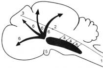
When developing the potential of action, a change in tissue excitability occurs, and this change proceeds but phases (Fig. 2). The state of the initial polarization of the membrane, which reflects the diaphragmal potential of rest, corresponds to the initial state of its excitability and, therefore, the cells are a normal level of excitability. During the period, the excitability of the tissue is improved, this phase of excitability was named primary exaltation. During the development of preparation, the membrane potential of rest approaches the critical level of depolarization and to achieve the latter, the strength of the irritant is less than the threshold (retaining).
During the development of spike (peak potential) there is avalanche-like admission of sodium ions inside the cell, resulting in recharging membrane and it loses the ability to respond to excitement to stimuli even overclocking force. This phase of excitability was named absolute refractoriness (absolute non-confidence). It lasts until the end of the recharge of the membrane. Absolute refractory, i.e., the complete non-responsibility of the membrane arises due to the fact that sodium channels are fully opened at the beginning, and then inactivated.
After the phase of recharging the membrane, the excitability is gradually restored to the initial level - phase relative refractoriness. It continues until the charge of the membrane is restored to the value corresponding to the critical level of depolarization. Since during this period, the membrane potential of rest is not restored, then the excitation of the tissue is lowered and the new excitement may occur only under the action of a super positioning stimulus. Reducing excitability in the phase of relative refractiveness is associated with partial inactivation of sodium channels and the activation of potassium.
The period of negative trace capacity corresponds to an increased level of excitability - phase of secondary exaltation. Since the membrane potential in this phase is closer to the critical level of depolarization, but compared with the state of chambers (initial polarization), then the irritation threshold is reduced, i.e., excitability is raised. In this phase, the new excitement may occur under the action of stimuli of the porch force. Sodium channels in this phase are inactivated by incompleteness. During the development of the positive trace potential, the causity of tissue is lowered - phase of secondary refractoriness. In this phase, the membrane potential increases (the state of hyperpolarization of the membrane), removing from the critical level of depolarization, the irritation threshold increases and the new arousal may occur only under the action of non-position irritants. The hyperpolarization of the membrane develops as a result of three reasons: first, the continuing output of potassium ions; secondly, the discovery, possibly channels for chlorine and the flow of these ions in the cytoplasm of the cell; Third, the strengthened work of the sodium-potassium pump.
4) (Cutting on nervous fibers)
The mechanism for the propagation of excitation among various nervous fibers of non-refinery. According to modern ideas, the spread of nervous fiber excitation is carried out on the basis of ionic mechanisms for generating the potential of action.
When the excitation of the incommable nerve fiber is local electric currents, which arise between its excited plot, charged negatively, and unexcited, charged positively, cause membrane depolarization to a critical level, followed by the generation of PD in the nearest point of the unexcited membrane. This process is repeated multiple times. Throughout the nervous fiber, the process of reproduction of the new PD at each point of the fiber membrane is occur. You call such an excitation and continuous.
The presence of a shell-haul of myelin fibers with high electrical resistance, as well as fiber areas devoid of shells (Ravier interceptions) Create conditions for a qualitatively new type of excitation on myelin nerve fibers. Local electric currents occur between the adjacent interceptions of Ranvier, since the membrane of the excited interception becomes charged negatively relative to the surface of the adjacent unexcited interception. These local currents are depolarized by the membrane of an unexcited interception before the critical level and the PD arises in it (Fig. 4). Consequently, the excitation seems to be "jumping" through the areas of nerve fibers, coated with myelin, from one interception to another. Such a mechanism for the spread of excitation is called salta or scrolling. The rate of such a method of excitation is significantly higher and it is more economical compared to the continuous excitation, since not the entire membrane is involved in the state of activity, but only its small areas in the field of interceptions.
Fig. 4. Scheme of excitation propagation in miserable (A) and myeline (b) nerve fibers.
The "jumping" of the potential of action through the site between interceptions is possible because the amplitude of PD is 5-6 times higher than the threshold value necessary to excite the adjacent interception. PD can "jump" not only through one, but also through two interpersonal gaps. This phenomenon can be observed by reducing the excitability of the neighboring interception under the action of any pharmacological substance, for example, novocaine, cocaine, etc.
Nervous fibers possess lability - the ability to reproduce a certain number of excitation cycles per unit of time in accordance with the rhythm of existing irritants. The measure of lability is the maximum number of excitation cycles, which can reproduce the nerve fiber per unit of time without transformation of the irritation rhythm. The lability is determined by the duration of the peak of the potential of the action, i.e. the phase of absolute refractoriness. Since the duration of absolute re-facterity in the spike potential of the nerve fiber is the shortest, the lability is the highest. Nervous fiber can reproduce up to 1000 pulses per second.
N. E. Vvedensky found that if the nerve site is subjected alteration (i.e., the effects of the damaging agent) by means of, for example, poisoning or damage, the lability of such a plot decreases sharply. Restoring the initial state of the nervous fiber after each action potential in the damaged area is slow. Under action to this section of frequent stimuli, it is not able to reproduce the specified irritation rhythm, and therefore pulses are blocked. Such a state of reduced lability was named by N. E. Vvedensky parabital. In the development of the state of parabiasis, there may be three, consistently replacing each other, phases: equalizing, paradoxical, brake.
IN equation phase The adjustment of the response to frequent and rare stimuli is occurring. In normal conditions of functioning of the nerve fiber, the magnitude of the response of the muscular fiber inexvurbed to them is obeys the law of force: the response is less for rare stimuli, and more irritants are more. Under the action of a parabiotic agent and with a rare irritation rhythm (for example, 25 Hz), all excitation impulses are carried out through a parabiotic portion, since the excitability after the previous pulse has time to recover. With a high rhythm of irritation (100 Hz), subsequent impulses can act at the moment when the nerve fiber is still in a state of relative refractority caused by the previous potential of action. Therefore, part of the pulses is not carried out. If only every fourth excitation (i.e. 25 pulses out of 100) are performed, then the amplitude of the response becomes the same as the rare stimuli (25 Hz) - the response is equalized.
IN paradoxical phase There is a further decrease in lability. At the same time, the response occurs on rare and frequent stimuli, but it is much smaller to frequent stimuli, since frequent stimuli reduce the lability, lengthening the absolute pulp phase. Therefore, there is a paradox - on rare stimuli, the response is more than frequent.
IN brake phase The lability is reduced to such an extent as rare, and frequent stimuli do not cause a response. At the same time, the membrane of the nerve fiber depolarized and does not go into the stage of repolarization, i.e., its initial state is not restored.
Parabiosis phenomenon underlies drug-based local anesthesia. The effect of anesthetic substances is also associated with a decrease in lability and disruption of the mechanism of excitation by nerve fibers.
Parabites - reversible phenomenon. If a parabiotic substance acts long, then after the cessation of its operation, the nerve comes out of the state of parabiosis through the same phases, but in the reverse order.
The mechanism for the development of the parabiotic state is reduced to the following. When exposed to parabiotic factor nervous fiber, the membrane ability to increase sodium permeability in response to irritation is disturbed. In the alteration site, the inacultation of sodium channels caused by the damaging agent is summed up with inactivation caused by a nervous impulse, and excitability decreases so much that the next pulse is blocked.
5) (Synapses, their types, structural features)
Physiology of synapses.
In the central nervous system, nervous cells are connected with each other by synapses. Sinaps. - It is a structural functional education that provides excitation or braking from nerve fibers to an innervized cell.
Sinapsy localization They are divided into central (located within the central nervous system, as well as in the ganglia of the vegetative nervous system) and peripherals (located outside the central nervous system, provide communication with cells of innervated tissue).
In functional attitudesinapses are divided by excitingin which, as a result of depolarization of the postsynaptic membrane, an exciting postsynaptic potential is generated, and brakesIn the presynaptic terminations of which the mediator is distinguished, a hyperpolarizing postsynaptic membrane and causing the occurrence of braking postsynaptic potential.
By the transmission mechanism Synapses are divided into chemical and electric. Chemical synapses transmit excitation or braking due to special substances - mediators. In action from the type of mediator Chemical synapses are divided into:
1. Holieregic (Mediator - Acetylcholine)
2. Adrenergic (Mediators - Adrenaline, Noradrenalin)
By anatomical classification Synapses are divided into neurosecretory, neuromuscular and inter-line.
Sinaps. Consists of three main components:
1. Presinautical membrane
2. Postsynaptic membrane
3. Synaptic slit
The presynaptic membrane is the end of the nervous cell process. Inside the process in the immediate vicinity of the membrane, there is a cluster of bubbles (granules) containing one or another mediator. Bubbles are in constant motion.
The postsynaptic membrane is part of the cell membrane of innervated tissue. Postsynaptic membrane, unlike the presexpotic protein chemoreceptors to biologically active (mediators, hormones), medicinal and toxic substances. An important feature of postsynaptic membrane receptors is their chemical specificity, i.e. The ability to join biochemical interaction only with a certain type of mediator.
The synaptic slit is the space between the pre- and postsynaptic membranes filled with a liquid close in composition to the blood plasma. Through it, the mediator slowly diffuses from the presynaptic membrane to postsynaptic.
The features of the structure of neuromuscular synapse cause it physiological properties.
1. One-sided excitation (from the provision of postsynaptic membrane), due to the presence of receptor-sensitive receptor mediator only in the postsynaptic membrane.
2. The synaptic delay in the excitation (time between the arrival of the pulse into the presynaptic ending and the beginning of the postsynaptic response) associated with a low rate of diffusion of the mediator into the synaptic slot compared with the speed of the pulse through the nerve fiber.
3. Low lability and high fatigue of synapse, due to the time of propagation of the previous pulse and the presence of absolute period period.
4. High electoral sensitivity of synapse to chemicals due to the specificity of the hemoreceptors of the postsynaptic membrane.
Stages of synaptic transmission.
1. Synthesis of mediator. In the cytoplasm of neurons and nerve endings, chemical mediators are synthesized - biologically active substances. They are constantly synthesized and deposited in the synaptic bubbles of nerve endings.
2. Secretion of the mediator. The release of the mediator from synaptic bubbles has a quantum character. In a state of rest, it is insignificant, and under the influence of the nerve impulse is sharply intensified.
3. Mediator interaction with postsynaptic membrane receptors. This interaction consists in selective changes in the permeability of the ion-selective effects of the effector cell in the field of active binding centers with the mediator. The interaction of the mediator with its receptors can cause excitation or inhibition of neuron, the reduction of muscle cells, the formation and separation of hormones by secretory cells. In the event of an increase in the permeability of sodium and calcium channels, the receipt of Na and Ca in the cell is increasing, followed by depolarization of the membrane, the occurrence of PD and the further transmission of the nervous pulse. Such synapses are called exciting. If the permeability of potassium channels and chlorine channels increases, there is an excess yield to from a cell with simultaneous diffusion into it cl, which leads to hyperpolarization of the membrane, a decrease in its excitability and development of braking postsynaptic potentials. The transfer of nerve impulses is hampered or completely stopped. Such synapses are called brakes.
Receptors interacting with AH are called cholinoreceptors. In functionality, they are divided into two groups: M - and n-cholinoreceptors. In synapses of skeletal muscles there are only n-cholinoreceptors, whereas in the muscles of internal organs are predominantly M-cholinoreceptors.
Receptors interacting with on, are called adrenoreceptors. In functionality, they are divided into alpha and beta-adrenoreceptors. In the postsynaptic membrane of smooth muscle cells of internal organs and blood vessels, both types of adrenoreceptors are often adjacent. The action on is depolarizing, if it interacts with alpha-adrenoreceptors (reducing the muscular shell of the walls of blood vessels or intestines), or brakes - when interacting with beta-adrenoreceptors (relaxation them).
4. Inactivation of the mediator. Inactivation (full loss of activity) The mediator is necessary to repolarize the postsynaptic membrane and restore the initial level of MP. The most important way to inactivation is hydrolytic splitting with inhibitors. For ah inhibitor is cholinesterase, for on and adrenaline - monoamine oxidase and catechoxymethyltransferase.
Another way to remove the mediator from the synaptic slit is "reverse capture" of its presynaptic endings (pinocytosis) and return axon transport, especially pronounced for catecholamines.
The coordination activities of the CNS lies the interaction of the processes of initiation and braking.
Excitation - This is an active process, which is a response of tissue for irritation and characterized by an increase in tissue functions.
Braking - This is an active process, which is a tissue response to irritation and characterized by a decrease in tissue functions.
Primary braking in the CNS occurs due to brake neurons. This is a special type of insertion neurons, which, when transmitting the pulse, highlight the brake mediator. Two types of primary braking are distinguished: postsynaptic and presynaptic.
Postsynaptic braking It occurs if the axon of the brake neuron forms a synaps with a neuron body and, highlighting the mediator, causes the cell membrane hyperpolarization, braking the cell activity.
Presinautical braking It occurs when the axon of the brake neuron forms a synaps with a axon of an exciting neuron, preventing the pulse.
6) (Spinal cord, its functions, participation in the regulation of muscle tone)
The spinal cord performs reflex and conductive functions. The first is provided by its nervous centers, the second conductive paths.
It has a segmental structure. Moreover, division into segments is functional. Each segment forms the front and rear roots. Rear are sensitive, i.e. Afferent, front engine, efferent. This pattern is called Bella Majandi law. The roots of each segment are innervating 3 metal components, but as a result of the overlap, each metaker is innervated by three segments. Therefore, with the damage to the front roots of one segment, the motor activity of the corresponding metaer is only weakened.
The morphologically body of the neurons of the spinal cord form its gray substance. It is functionally all of its neurons are divided into motioneons, inserts, neurons of the sympathetic and parasympathetic departments of the vegetative nervous system. Motionic, depending on the functional value, are divided into alpha - and gamma-motoneurons. The fibers of the afferent paths are coming to A-motor mechanons, which begin with intrafusal, i.e. receptor muscle cells. The bodies of the A-motorhones are located in the front horns of the spinal cord, and their axons innervate skeletal muscles. Gamma-motoneurons regulate the voltage of muscle spindles i.e. Intrafusal fibers. Thus, they are involved in the regulation of reducing skeletal muscles. Therefore, when the front roots are cut, the muscular tone disappears.
Innexers provide a link between the spinal cord centers and the overlying CNS departments.
The neurons of the sympathetic department of the vegetative nervous system are in the lateral horns of the chest segments, and the parasympathetic in the sacrats department.
The conductor function consists in ensuring communication of peripheral receptors, the centers of the spinal cord with the overlying sections of the CNS, as well as its nervous centers among themselves. It is carried out by conductive paths. All the paths of the spinal cord are divided into their own or propropinyl, ascending and descending. Propropinal paths are associated with each other nervous centers of different spinal cord segments. Their function is to coordinate the tone of muscles, movements of various body meta chairs.
Rising paths include several paths. Gulf beams and bourdaches carry out nerve impulses from muscle proprigororeceptors and tendons to the appropriate centers of the oblong brain, and then Talamus and the somatosensory cortex zones. Thanks to these paths, an assessment and correction of the position of the body is made. The beams of Govers and Flexig transmit excitation from the proprigororeceptors, the mechanoreceptors of the skin to the cerebellum. This ensures perception and unconscious coordination of postures. The spinctalamatic paths spend signals from pain, temperature, tactile skin receptors to the thalamus, and then somatosensory cortex zones. They ensure the perception of the corresponding signals and the formation of sensitivity.
The downstream paths are also formed by several paths. Corkospinal paths go from pyramid and extrapyramidal cortex neurons to the A-motor-mechanons of the spinal cord. Due to them, regulation of arbitrary movements is carried out. Rubrosspinal path spends signals from the red core of the middle brain to A-motor mechanics of the muscles of flexors. The vestiblympinal path transmits signals from the vestibular cores of the oblong brain, primarily the dealers kernel, to the A-motor mechanons of the muscle of the extensors. Due to these two paths, the tone of the corresponding muscles is regulated when the body position is changed.
All spinal cord reflexes are divided into somatic, i.e. Motor and vegetative. Somatic reflexes are divided into tendon or mitatic and skin. Tendor reflexes occur in mechanical irritation of muscles and tendons. Their slight stretching leads to the excitation of tendon receptors and the a-motor meters of the spinal cord. As a result, there is a reduction in muscles, first of all the extensors. The tendon reflexes include the knee, achilles, elbow, bruside, etc., arising from mechanical irritation of the corresponding tendons. For example, the knee is the simplest monosynaptic, since only one synaps in its central part. Skin reflexes are caused by irritation of skin receptors, but manifested by motor reactions. They are the sole and abdominal (explanation). Spinal nerve centers are under the control of the overlying. Therefore, after cuts between the oblong and spinal cord, the spinal shock arises and the tone of all muscles will significantly decrease.
Vegetative spinal cord reflexes are divided into sympathetic and parasympathetic. Those and others are manifested by the reaction of internal organs for irritation of skin receptors, internal organs, muscles. The vegetative neurons of the spinal cord form lower centers for the regulation of the tone of vessels, cardiac activity, the lumen of the bronchi, sweating, urinary, defecation, erection, eiyaculation, etc.
7) (The oblong brain and bridge, their functions, participation in the regulation of muscle tone)
Medulla
Features of the functional organization. An oblong brain (Medullaoblongata) in a person has a length of about 25 mm. It is a continuation of the spinal cord. Structurally on the diversity and structure of the cores, the oblongable brain is more complicated than the dorsal. In contrast to the spinal cord, it does not have a metairene, repeated structure, the gray substance in it is not located in the center, but the cores to the periphery.
In the oblong brain there are olives associated with the spinal cord, an extrapyramine system and cerebellum - this is a subtle and wedge-shaped nucleus of proprioceptive sensitivity (the core of Gully and Burdach). Here are the crossings of descending pyramid pathways and rising paths formed by thin and wedge-shaped beams (goals and burgunda), a reticular formation.
The oblongable brain at the expense of its nuclear formations and the reticular formation is involved in the implementation of vegetative, somatic, taste, auditory, vestibular reflexes. The peculiarity of the oblong brain is that its kernels, excitement consistently, ensure the performance of complex reflexes that require consistent inclusion of different muscle groups, which is observed, for example, when swallowing.
The cores of the following cranial nerves are located in the oblong brain:
a pair of VIII cranial nerves - the predvevno-ulitskoy nerve consists of snippening and finally parts. Sniply kernel lies in the oblong brain;
a pair of IX is a low-calorie nerve (p. Glossopharyngeus); Its core is formed by 3 parts - motor, sensitive and vegetative. The motor part participates in the innervation of the muscles of the pharynx and the oral cavity, sensitive - receives information from the receptors of the taste of the back third of the tongue; Vegetative innervates salivary glands;
a pair X - a wandering nerve (N.Vagus) has 3 cores: the vegetative innervates the larynx, the esophagus, heart, stomach, intestines, digestive glands; sensitive receives information from the receptors of the alveoli light and other internal organs and the motor (so-called mutual) provides a sequence of cutting muscles, larynx when swallowing;
pair Xi - additional nerve (N.Accessorius); Its kernel is partially located in the oblong brain;
a pair of XII - Podium-speaking nerve (N.Hypoglossus) is a motor nerve of the tongue, its core is mostly located in the oblong brain.
Sensory functions. The oblong brain regulates a series of sensory functions: the reception of the skin sensitivity of the face - in the touch nucleus of the trigeminal nerve; Primary analysis of the reception of taste - in the nucleus of the Language nerve; Reception of auditory irritation - in the kernel of the sniplike nerve; Reception of vestibular irritations - in the upper vestibular core. In the ass, deep, visceral sensitivity, part of which switches here on the second neuron (thin and wedge-shaped kernel) in the assays of the oblong brain. At the level of the continued brain, the listed sensory functions implement a primary analysis of the strength and quality of irritation, then the processed information is transmitted to subcortical structures to determine the biological significance of this irritation.
Explore functions. Through the continued brain pass all the ascending and descending paths of the spinal cord: the spinal thalamic, corticospinal, ruble. It takes the beginning of the vestiblospinal, olivospinal and reticulous-permanent paths, providing the tone and coordination of muscle reactions. In the oblong brain, paths from the cortex of the Big Brain - Corresponding Ways. The rising paths of proprioceptive sensitivity from the spinal cord are completed here: thin and wedge-shaped. Such cerebral formations, like a bridge, middle brain, cerebellum, Talamus, hypothalamus and large brain bark, have bilateral connections with an oblong brain. The presence of these links indicates the participation of the oblong brain in the regulation of the tone of skeletal muscles, vegetative and higher integrative functions, the analysis of sensory irritations.
Reflex features. Numerous reflexes of the oblong brain shall be divided into vital and insecificance, however, such a submission is sufficiently conditionally. The respiratory and vascular centers of the oblong brain can be attributed to vital centers, as they are closed by a number of heart and respiratory reflexes.
An oblong brain organizes and implements a number of protective reflexes: vomiting, chihana, cough, tears, a century closure. These reflexes are implemented due to the fact that the information about the irritation of the receptors of the mucous membrane of the eye, the oral cavity, the larynx, the nasopharynxes through the sensitive branches of the triangistic and language nerve falls into the core of the oblong brain, hence the team comes to the motor nucleus of a trigeminal, wandering, facial, language, supplementary Podium nerves, as a result, a particular protective reflex is implemented. In the same way due to the consistent inclusion of muscle groups of head, neck, chest and diaphragms, food behavior reflexes are organized: sucking, chewing, swallowing.
In addition, the oblong brain organizes the reflexes of maintaining poses. These reflexes are formed due to the afferentation from the receptors of the Thread Street and semicircular channels in the upper vestibular kernel; From here, reworked information evaluation of the need to change poses is sent to the lateral and medial vestibular nuclei. These cores are involved in the definition of what muscle systems, the spinal cord segments must take part in the change of poses, therefore, from neurons of the medial and lateral kernel in the vestiblospinal pathway, the signal comes to the front horns of the corresponding spinal cord segments innervating muscles, whose participation in changing poses in This moment is necessary.
Change the posture is carried out by static and static reflexes. Static reflexes regulate the tone of skeletal muscles in order to hold a certain position of the body. Stasinetic reflexes of the oblong brain ensure the redistribution of the tone of the muscles of the body to organize the posture corresponding to the moment of straight or rotational motion.
Most of the autonomous reflexes of the oblong brain are implemented through the kernel of the vagus nerve, which receive information about the state of the heart, vessels, digestive tract, light, digestive glands, etc. In response to this nucleus information, the nucleus and secretory reactions of these organs organize.
The excitation of the wandering nerve nuclei causes strengthening the reduction of the smooth muscles of the stomach, intestines, gallbladder and at the same time relaxing the sphincters of these organs. At the same time, the work of the heart slows down and weakens, the lumen of the bronchi is narrowed.
The activity of the wandering nerve nuclei is also manifested in strengthening the secretion of bronchial, gastric, intestinal glands, in the excitation of pancreas, secretory liver cells.
In the oblong brain, the center of salivation is localized, the parasympathetic part of which ensures the strengthening of general secretion, and the sympathetic - protein secretion of the salivary glands.
The structure of the reticular formation of the oblong brain is located respiratory and vascular centers. The peculiarity of these centers is that their neurons are able to be excited reflexively and under the influence of chemical stimuli.
The respiratory center is localized in the medial part of the reticular formation of each symmetric half of the oblong brain and is divided into two parts, inhalation and exhalation.
In the reticular formation of the oblong brain, another vital center is presented - a vasomotor center (regulation of vascular tone). It functions in conjunction with the overlying structures of the brain and primarily with the hypothalamus. The excitation of the vasomotory center always changes the rhythm of breathing, the tone of the bronchi, the muscles of the intestine, the bladder, the ciliary muscle, etc. This is due to the fact that the reticular formation of the oblong brain has synaptic ties with the hypothalamus and other centers.
In the middle deposits of the reticular formation are neurons that form a reticulospinal path, which has a braking effect on the spinal cord motoneurons. At the bottom of the IV ventricle there are neurons of the Blue Spot. Their mediator is norepinephrine. These neurons cause activation of the reticulospinal flow to the "fast" sleep phase, which leads to the braking of spinal reflexes and a reduction in muscle tone.
Symptoms of damage. Damage to the left or right half is oblong brain above the crossroads of the rising paths of propriceceptive sensitivity causes the side of damage to the sensitivity and work of the muscles of the face and head. At the same time, on the opposite side with respect to the side of the damage, skin sensitivity disorders and motoric parameters of the body and limbs are observed. This is due to the fact that the ascending and descending conducting pathways from the spinal cord and in the spinal cord are crossed, and the core nerve kernels innervate their half of the head, i.e., the cranial nerves do not intersect.
Bridge
The bridge (PonscereBri, PonSvarolii) is located above the oblong brain and performs sensory, conductive, motor, integrative reflex functions.
The bridge includes a facial, trigeminal, discrepancing, predvevno-snelled nerve (vestable and snellest kernels), the kernel of the pre-propelled part of the sentence-snelled nerve (vestibular nerve): lateral (deuteris) and top (behtereva). The reticular formation of the bridge is closely related to the reticular formation of medium and oblong brain.
An important structure of the bridge is the middle leg of the cerebellum. It is it that provides functional compensatory and morphological connections of a large brain cortex with cerebellum hemispheres.
The sensory functions of the bridge are provided by the nuclei of the sentence-snelled, trigeminal nerves. Ulitkaya part of the predver-snellest nerve ends in the brain in the snellers; The finver part of the sentence-snelled nerve is in the triangular kernel, the dealers core, the Bekhterev core. Here is a primary analysis of vestibular irritation of their strength and orientation.
The sensitive kernel of a trigeminal nerve receives signals from the facial skin receptors, the front departments of the scalp, the mucous membrane of the nose and mouth, teeth and the conjunctivation of the eyeball. Facial nerve (p. Facialis) innervates all the facial muscles. An abduction nerve (p. Abducans) innerves the straight lateral muscle, the discharge of the eyeball of the duck.
The motor portion of the trigeminal nerve nucleus (PRIGEMINUS) innervates the chewing muscles, the muscle that pulls the drumpoint, and the muscle pulling the sky curtain.
Conductive bridge function. Provided by longitudinally and transversely arranged fibers. The transversely arranged fibers form the upper and lower layers, and between them goes from the cortex of the big brain the pyramidal paths. Between the transverse fibers are neural clusters - the bridge cores. From their neurons begin transverse fibers, which go to the opposite side of the bridge, forming the middle leg of the cerebellum and ending in its crust.
In the bridge covers, there are longitudinally running bunches of the fibers of the medial loop (LemniscusMedialis). They intersect transversely walking fibers of the trapezoid body (Corpustrapezoideum), which are axons of the street part of the sentence-snelled nerve of the opposite side that end in the top olive kernel (Olivasuperior). From this core goes the side loop path (Lemniscuslateralis), which are sent to the back of the mid-brain and the medial crankshafts of the intermediate brain.
In the brain coated, the front and rear kernel of the trapezoid body and the lateral loop are localized. These kernels together with the upper olive provide a primary analysis of information from the hearing organ and then transmit information to the rear bumps of Quarters.
The covers also contains a long medial and textospinal path.
Own neurons of the structure of the bridge structure form its reticular formation, the kernel of the facial, discharge nerve, motor portion of the nucleus and the average sensory core of the trigeminal nerve.
The reticular formation of the bridge is the continuation of the reticular formation of the oblong brain and the beginning of the same mid-brain system. The axons of the neurons of the reticular formation of the bridge go to the cerebellum, in the spinal cord (reticulospinal path). The latter activate the neurons of the spinal cord.
The reticular formation of the bridge affects the bark of a large brain, causing its awakening or a sleepy state. In the reticular formation of the bridge there are two groups of cores that belong to the overall respiratory center. One center activates the center of the breath of the oblong brain, the other - the center of the exhalation. The neurons of the respiratory center, located in the bridge, adapt the operation of respiratory cells of the oblong brain in accordance with the changing condition of the body.
8) (Medium brain, its functions, participation in the regulation of muscle tone)
Morphofunctional organization. The middle brain is presented by four-headed and legs of the brain. The largest nuclei of the middle brain is the red core, a black substance and core core (oxotiac and block) nerves, as well as the kernel of the reticular formation.
Sensory functions. Implemented by receipt of visual, auditory information.
Explore function. It is that through it all the rising paths to the overlying Talamus (medial loop, spiinolamic path), a large brain and cerebellum. The descending paths go through the middle brain to the oblong and spinal cord. This is a pyramid path, cortical-bridge fibers, a ruler-global path.
Motor function. It is implemented at the expense of the nucleus of the block nerve (N. Trochlearis), the nuclei of the Oculomotor nerve (oculomotorius), the red kernel (Nucleusruber), the black substance (substantianigra).
The red cores are located at the top of the brain legs. They are associated with a big brain bark (descending from the crust of the path), subcortical nuclei, cerebellum, spinal cord (red-cerebral pathway). Basal ganglia brain, cerebellum have their endings in red nuclei. Violation of the links of red nuclei with the reticular formation of the oblong brain leads to decependence rigidity. This condition is characterized by a strong voltage of muscle-extensor extremities, neck, back. The main reason for the occurrence of decependence rigidity is the pronounced activating effect of the lateral vestibular nucleus (the actor of the deuteris) on the vestibule motoneurons. This is the effect of the maximum in the absence of the braking effects of the red nucleus and the overlying structures, as well as the cerebellum. When the brain is cut below the kernel of the lateral vestibular nerve, decerebraction rigidity disappears.
Red nuclei, receiving information from the motor zone of the large brain cortex, subcortical nuclei and cerebellum about the preparing movement and state of the musculoskeletal system, send corrective pulses to the spinal cord motnelones over a rubry-block path and thereby regulate the muscles tone, preparing its level to the outlined arbitrary movement. .
Another functionally important core of the mid-brain is a black substance - located in the legs of the brain, regulates the acts of chewing, swallowing (their sequence), ensures accurate movements of the fingers of the brush hand, for example, when writing. The neurons of this kernel are able to synthesize the mediator dopamine, which is supplied by axonal transport to the basal ganglia of the brain. The damage to the black substance leads to a violation of the plastic tone of the muscles. The fine regulation of the plastic tone when playing the violin, writing, performing graphic works is provided by black substance. At the same time, with a long retention of a certain posture, plastic changes in the muscles occur due to changes in their colloidal properties, which ensures the smallest energy costs. Regulation of this process is carried out by black substance cells.
Neurons of the nuclei of the glasses and block nerves regulate the movement of the eye up, down, outward, to the nose and down to the corner of the nose. The neurons of the additive kernel of the icy nerve (Yakubovich's kernel) regulate the lumen of the pupil and the crystal curvature.
Reflex features. Functionally independent structures of the middle brain are the bugs of four. The top of them are primary subcortex centers of the visual analyzer (together with the lateral crankshafts of the intermediate brain), the lower - auditory (together with the medial crankshafts of the intermediate brain). They occur in the primary switching of visual and hearing information. From the bugs, four axons of their neurons go to the reticular formation of the barrel, the spinal cord motnelones. Thorough neurons can be polymodal and detector. In the latter case, they react only to one sign of irritation, for example, the change of light and darkness, the direction of the movement of the light source, etc. The main function of the buggers is four-headed - the organization of the alarming reaction and the so-called start-reflexes for sudden, not yet recognized, visual or sound Signals. The activation of the middle brain in these cases through the hypothalamus leads to an increase in muscle tone, the increase in heart cuts; There is preparation for avoiding, to a defensive reaction.
Quarterly organizes approximate visual and auditory reflexes.
A person has a quirky reflex is a watchdog. In cases of increased rapid excitability with sudden sound or light irritation, a person has shuddered, sometimes jumping to feet, screaming, the most rapid removal from the stimulus, sometimes unrestrained.
In case of violation of a four-champion reflex, a person cannot quickly switch from one type of movement to another. Consequently, quadruses take part in the organization of arbitrary movements.
Reticular brain stem formation
The reticular formation (Formatioreticularis; of the Russian Federation) of the brain is represented by a neuron network with numerous diffuse bonds among themselves and practically with all the structures of the central nervous system. The Russian Federation is located in the thickness of the gray matter of the oblong, middle, intermediate brain and is initially connected with the Russian Federation of the spinal cord. In this regard, it is advisable to consider it as a unified system. Network links of the neurons of the Russian Federation have allowed the dealers to call it the reticular formation of the brain.
The Russian Federation has direct and inverse relationships with a large brain bark, basal ganglia, intermediate brain, cerebellum, medium, oblong and spinal cord.
The main function of the Russian Federation is the regulation of the level of activity of the large brain cortex, cerebellum, thalamus, spinal cord.
On the one hand, the generalized nature of the influence of the Russian Federation on many brain structures gave reason to consider it a non-specific system. However, studies with irritation of the Russian Federation trunk showed that it can selectively provide an activating or inhibitory effect on different forms of behavior, on sensory, motor, visceral brain systems. The network structure ensures high reliability of the functioning of the Russian Federation, resistance to damaging effects, as local damage is always compensated by the preserved network elements. On the other hand, the high reliability of the functioning of the Russian Federation is ensured by the fact that the irritation of any of its parts is reflected on the activity of the entire Russian Federation of this structure due to the diffusion of connections.
Most of the neurons of the Russian Federation has long dendrites and a short axon. There are giant neurons with a long axon that form paths from the Russian Federation to other areas of the brain, for example, in the descending direction, reticulous and rubn. The axons of the neurons of the Russian Federation form a large number of collaterals and synapses, which are eared on the neurons of various brain departments. The axons of the neurons of the Russian Federation, going to the bark of a large brain, end here on dendrites I and II of the layers.
The activity of the RF neurons is different and in principle, similar to the activity of neurons of other brain structures, but among the neurons of the Russian Federation there are those that have sustainable rhythmic activity that does not depend on the incoming signals.
At the same time, in the Russian Federation of the middle brain and the bridge there are neurons, which are silent "silent", i.e., impulses are not generated, but are excited when stimulating visual or auditory receptors. These are the so-called specific neurons, providing a quick response to sudden, unidentified signals. A significant number of neurons of the Russian Federation are polyessens.
In the Russian Federation oblong, mid-brain and bridge convert signals of various sensory. On the neurons of the bridge come signals mainly from somatosensory systems. Signals from visual and hearing sensory systems mainly come on the neurons of the middle brain.
The Russian Federation controls the transmission of sensory information going through the thalamus kernels due to the fact that with intensive external irritation of neurons of nonspecific thalamus nuclei slow down, thereby removing their inhibitory effect from the relay nuclei of the same Talamus and facilitates the transmission of sensory information in the bark of a large brain.
In the Russian Federation, the bridge, oblong, mid-brain there are neurons that react to pain irritations coming from muscles or internal organs, which creates a common diffuse discomfort, not always clearly localized, painful feeling of "stupid pain."
The repetition of any type of stimulation leads to a decrease in the pulse activity of the neurons of the Russian Federation, that is, the processes of adaptation (addiction) are inherent in the neurons of the Russian Barrel of the Brain.
The Russian brain trunk is directly related to the regulation of the muscular tone, since the stem of the brain is received signals from visual and vestibular analyzers and cerebellum. From the Russian Federation to the motor mechanons of the spinal cord and the clerk nerve nuclei, signals coming the position of the head, the body, etc.
Reticular pathways that facilitate the activity of motor spinal cord systems originate from all departments of the Russian Federation. The paths coming from the bridge inhibit the activity of the spinal cord motioneons, innervating flexor muscles, and activate the motionones of the muscle extensors. The paths coming from the Russian Federation of the oblong brain cause opposite effects. Irritation of the Russian Federation leads to tremor, increase muscle tone. After the termination of irritation, the effect caused by it is preserved for a long time, apparently due to circulation of excitation in the neuron network.
The brain barrel participates in the transfer of information from the cortex of a large brain, the spinal cord to the cerebellum and, on the contrary, from the cerebellum to the same systems. The function of these links is to prepare and implement the motility associated with addiction, indicative reactions, painful reactions, the organization of walking, eye movements.
Regulation of the vegetative activities of the Russian Federation is described in section 4.3, here we note that this regulation is shown in the functioning of respiratory and cardiovascular centers. In the regulation of vegetative functions, the so-called starting neurons of the Russian Federation have great importance. They give rise to the circulation of excitation inside the neurons group, providing a tone of adjustable vegetative systems.
The influence of the Russian Federation can be divided in general on descending and ascending. In turn, each of these influences has a braking and exciting effect.
The ascending influences of the Russian Federation on the bark of a large brain increase its tone, regulate the excitability of its neurons, without changing the specifics of the answers to adequate irritation. The Russian Federation affects the functional state of all sensory areas of the brain, therefore, it matters in integrating sensory information from different analyzers.
The Russian Federation is directly related to the regulation of cycle wake-sleep cycle. Stimulation of some structures of the Russian Federation leads to the development of sleep, the stimulation of others causes an awakening. Magong and D. Morutski put forward a concept that all types of signals coming from peripheral receptors are reached by the collateral of the Russian Federation of the oblong brain and the bridge, where they switch to neurons that give rising paths to the Talamus and then in the bark of the big brain.
The excitement of the Russian Federation of the oblong brain or the bridge causes synchronization of the activity of the large brain cortex, the appearance of slow rhythms in its electrical indicators, sleepy braking.
The excitation of the middle of the brain causes the opposite effect of awakening: the desynchronization of the electrical activity of the cortex, the appearance of rapid low-amplitude β-like rhythms in the electroencephalogram.
G. Bremer (1935) showed that if you cut the brain between the front and rear beatings of Quirms, the animal ceases to respond to all types of signals; If the cut to produce between the oblong and middle brain (while the Russian Federation retains a bond with the front brain), the animal reacts to light, sound and other signals. Consequently, maintaining the active analyzing state of the brain is possible when conservation of communication with the front brain.
The activation reaction of the large brain cortex is observed in the irritation of the Russian Federation of oblong, medium, intermediate brain. At the same time, irritation of some Talamus nuclei leads to the emergence of limited local excitation sites, and not to the general excitation, as it happens when irritating other departments of the Russian Federation.
The brain trunk can have not only an exciting, but also a braking effect on the activity of the cortex of the brain.
The descending effects of the Russian brain trunk on the regulatory activities of the spinal cord were established by I. M. Sechenov (1862). It was shown that with the irritation of the middle brain, salt crystals at the frog, the reflexes of the swelling of the paws occur slowly, require stronger irritation or do not appear at all, i.e. are braking.
Magong (1945-1950), causing local irritations to the Russian Federation of the oblong brain, found that when annoying some points are inhibited, becoming sluggish reflexes of bending the front paw, knee, cornea. When irritating the Russian Federation at other points of the oblong brain, the same reflexes were lighted easier, they were stronger, that is, their implementation was facilitated. According to Maguna, the brake influences on the spinal cord reflections can only have an oblong brain of the Russian Federation, and facilitating influences are regulated by the entire RF barrel and spinal cord.
9) (Cerebellum, his participation in the regulation of motor and vegetative functions)
Cerebellum (Cerebellum, small brain) is one of the integrative structures of the brain, participating in the coordination and regulation of arbitrary, involuntary movements, in the regulation of vegetative and behavioral functions.
Features of the morphofunctional organization and communication of the cerebellum. The implementation of these functions is ensured by the following morphological features of the cerebellum:
1) The cerebel bark is built quite similarly, has stereotypical communications, which creates conditions for rapid information processing;
2) the main neural element of the cortex - the Purkinier cell, has a large number of inputs and forms the only axes output from the cerebellum, the collateral of which ends on nuclear structures;
3) Purkinier cells are projected by almost all species stimuli: propriceceptive, skin, visual, auditory, vestibular, etc.;
4) Outputs from the cerebellum provide its connection with the cortex of a large brain, with stem formations and spinal cord.
The cerebellum is anatomically and is functionally divided into old, ancient and new parts.
To the old part of the cerebellum (Archicerebellum) - the vestibular cerebellum - refers to a flock-floccular share. This part has the most pronounced links with the vestibular analyzer, which explains the value of the cerebellum in the regulation of equilibrium.
The ancient part of the cerebellum (PaleoCereebellum) is a spinal cerebellum - consists of sections of the worm and pyramid of the cerebellum, tongue, a nearby department and gets information mainly from proprodoretic muscle systems, tendons, periosteum, joint shells.
The new cerebellum (neocereebellum) includes a bark of cerebellum hemispheres and sections of the worm; It receives information from the cortex, mainly in the frontal-brother-in-law path, from visual and auditory recipe systems, which indicates its participation in the analysis of visual, auditory signals and the organization of the reaction on them.
The cerebulic cortex has a specific, nowhere in the CNS is not repeated, the structure. The top (i) layer of cerebellum core is a molecular layer consists of parallel fibers, branches of dendrites and axons of the II and III layers. In the lower part of the molecular layer there are basket and star cells, which ensure the interaction of Purkin cells.
The middle (ii) layer of the crust is formed by Purkinier cells built in one row and having the most powerful dendritic system in the CNS. On the dendritic field of one cell Purkin can be up to 60,000 synapses. Consequently, these cells perform the task of collecting, processing and transmitting information. The axons of the Purkinier cells are the only way by which the cerebel bark transmits information in its kernel and the core of the large-brain structure.
Under the II layer of the crust (under the Purkin cell cells) lies with a granular (III) layer consisting of grain cells, the number of which reaches 10 billion axons of these cells rise up, T-figuratively divided on the surface of the crust, forming a path of contacts with Purkinier cells. Here are the Cells of Golges.
The information goes from the cerebellum through the upper and lower legs. Through the upper legs, signals go to the thalamus, in the bridge, the red core, the core of the brain, in the reticular formation of the mid-brain. Through the lower legs of the cerebellum, the signals go to the oblong brain to its vestibular nuclei, oliv, reticular formation. Medium legs of the cerebellum bind a new cerebellum with a frontal shared brain.
The pulsed neuron activity is recorded in the Purkinier cell layer and the granular layer, and the frequency of generation of pulses of these cells ranges from 20 to 200 per second. Cerezere kernel cells generate impulses significantly less often - 1-3 pulse per second.
Stimulation of the top layer of cerebellum core leads to a long (up to 200 ms) inhibition of the activity of Purkinier cells. The same braking occurs with light and sound signals. The total changes in the electric activity of the cerebellum cortex on the irritation of the sensitive nerve of any muscle look in the form of positive oscillation (braking of the cortex activity, hyperpolarization of Purkinier cells), which occurs after 15-20 ms and lasts 20-30 ms, after which the excitation wave is arising lasting up to 500 MS (depolarization of Purkiny cells).
In the ceremony of the cerebeller from the skin receptors, muscles, articular shells, the suspension signals are coming by so-called spinal cerebellar paths: on the rear (dorsal) and front (ventral). These paths to the cerebellum pass through the lower olive of the oblong brain. From the cells of Olive, the so-called climbing fibers come, which are branched on dendrites of Purkinier cells.
The core of the bridge is sent to the afferent paths into the cerebellum forming mossy fibers, which end in the cells - grains of the III cerebellum core layer. There is an afferent connection between the cerebellar and a bluish mid-brain place with adrenergic fibers. These fibers are able to diffusely throw away the norepinex in the intercellular space of the cerebeller's cortex, thereby humorly change the state of excitability of its cells.
A axons of cells of the III layer of cerebelic cerebral core cause the braking of Purkinier cells and the grain cells of their own layer.
Purkinier cells in turn hamper the neuron activity of the cerebellum kernels. The cerebel kernels have high tonic activity and regulate the tone of a number of motor intermediate, medium, oblong, spinal cord.
The cerebeller's subheading system consists of three functionally different nuclear formations: the cores of the tent, a plug, spherical and gear core.
The core of the tent receives information from the medial zone of cerebel bark and is associated with the core of the deuteris and the Russian Federation of oblong and mid-brain. Hence the signals go along the reticulous relief to the spinal cord motnelones.
The intermediate core of the ceremony is projected onto a plug and spherical core. Communication from them goes to the middle brain to the Red Kero, then in the spinal cord on the ruble path. The second way from the intermediate kernel goes to the Talamus and then in the motor zone of the large brain cortex.
The gentle core, receiving information from the lateral zone of the cerebel bark, is associated with the thalamus, and through it - with the motor zone of the large brain cortex.
Cereselic control of motor activity. Efferent cerebellum signals to the spinal cord regulate the power of muscle contractions, provide the ability to continuously tonic muscle reduction, the ability to maintain the optimal muscle tone alone or when driving, to commend random movements with the purpose of this movement, quickly move from bending to extension and vice versa.
The cerebellum provides synergies of cuts of different muscles in difficult movements. For example, making a step when walking, a person comes forward a foot, at the same time the center of gravity of the body is transferred forward with the participation of muscles of the back. In cases where the cerebellum does not perform its regulatory function, a person has disorders of motor functions, which is expressed by the following symptoms.
1) Asthenia (Astenia - weakness) - reduced muscle reduction force, fast muscle fatigue;
2) Astasia (Astasia, from Grech.a - not, Stasia - standing) - loss of ability to long-term muscle reduction, which makes it difficult for standing, sitting, etc.;
3) Dystonia (Distonia - a tone impairment) is an involuntary increase or decrease in muscle tone;
4) Tremor (Tremor - trembling) - trembling fingers, brushes, heads alone; This tremor is intensified when driving;
5) Distance (Dismetria is a violation of the measure) - disorder of uniformity of movements, expressed either in unnecessary or insufficient motion. The patient is trying to take the object from the table and hesitates the hand for the subject (hypertery) or it does not convey it to the subject (hypometry);
6) Ataxia (ATAKSIA, from Grech.A - denial, Taksia - order) - disruption of coordination of movements. Here, it is brighter that the impossibility of performing movements is in the desired order, in a certain sequence. The manifestations of ataxia are also adiadochokinesis, asiergia, drunk-shaky gait. With adiadochokine, a person is not able to quickly rotate the palms down-up. When muscle asyngia, he is not able to sit down from the position lying without help. Drunk gait is characterized by the fact that a person walks, spreading his legs widespread, staggering from the side away from the walking line. Congenital motor acts in humans are not so much (for example, sucking), most of the movements he learns during the life and they become automatic
- A number of nuclear structures occupying a central place in the stem of the brain. The morphological structure of the reticular formation is very similar to the mesh, and it was for this sign that first drew the attention of the German Anatom of Otto Daters. It was on this basis that he called this structure (lat. Reticulum - Mesh, Formatio - Education). I agree! "Mesh education" - sounds not as cool 🙂
Since the reticular formation passes through the entire brain barrel, it would be anatomically to divide it on the departments of the oblong brain, the Varoliev of the bridge and the middle brain, but since the individual parts of this structure are engaged in common things for them, then it is considered as a unified Structure.
To understand, I will give a comparison ... If you ever have been at concerts or at least watched them on TV, you probably noticed huge panels with a bunch of buttons, togglers, switches, etc. With these panels, the operator sets the quality of the sound, downstream and increasing others, as well as chromaticity, brightness, contrast, etc. So the reticular formation is engaged in this. That is, it receives signals absolutely from all downstream and rising paths, recycles them, produces new coordinating signals and gives them to the appointment, providing us with normal perception.
Some neurons R. f. Show background activity, discharged 5-10 times per second. These nervous centers affect the bark of the brain, constantly supporting consciousness in us. If these centers are destroyed, animals comes.
For comparison I will explain. The reticular formation supports consciousness in us as well as fire in the fire supports boiling water in the boiler. It is worth putting fire - and boiling water, as well as consciousness, ceases to show activity. Consequently, in R. f. There are one of the centers of sleep and wakefulness.
With the brain cortex in general, communication is special. It is clear that R.F. Responsible for and excitement in the core of the brain. The bark of the brain, in turn, also has brake and exciting influences on the reticular formation. By forming closed neural connections, these two systems mutually regulate each other and balance their influences.
conclusions
The functions of the reticular formation are not sufficient due to the high degree of complexity of the organization of this structure, but the available data is sufficient for the following conclusions:
It affects the level of consciousness by interacting in the bark of the brain. Participates in the cycle in giving emotional color sensory signals in the art. Pain, by carrying out afferent information to the limbic system. By mutual coordination of afferent and efferent systems involved in the formation of vital reflexes. Also takes part in the vegetative functions of the body and as an important component of the engine centers of the brain.
Reticular formation The network of neurons of various types and sizes that have numerous connections among themselves, as well as with all the structures of the CNS. It is located in the thickness of the gray substance of the oblong, middle and intermediate brain and regulates the level of functional activity (excitability) of all nerve centers of these CNS departments. In the same way, it affects the bark of large hemispheres.
Two subsystems that perform different organizing functions are distinguished into the CNS. specific and nonspecific. The first provides perception, conducting, analysis and synthesis of signals of specific sensitivity. These include all its types, i.e. visual, hearing, pain, etc.
Nonspecific The subsystem is a reticular formation. It has a generalized exciting or inhibitory effect on many brain structures. Consequently, it can regulate the level of functional activity of motor, sensory, visceral systems and the body as a whole. When the nerve impulses go on a specific conductive paths, they come to the collaterals of these paths to the neurons of the reticular formation. This leads to their diffuse excitation. From the neurons of the reticular formation, the excitation is transmitted to the bark, which is accompanied by the excitation of neurons of all its zones and layers. Due to this upward activating influence of the reticular formation, the activity of analytical synthetic activity increases, the speed of reflexes increases, the body is prepared for the reaction to an unexpected situation. Therefore, the reticular formation participates in the organization of defensive, sexual, digestive behavior. On the other hand, it can selectively activate or brake certain brain systems. In turn, the bark of large hemispheres, through descending paths, can have an exciting effect on the reticular formation.
The descending reticulospinal paths go from the reticular formation to the neurons of the spinal cord. Therefore, it can extend downward exciting and braking influences on its neurons. For example, its hypothalamic and mezanecephalous departments increase the activity of alpha-motoneurons of the spinal cord. As a result, a tone of skeletal muscles is growing, motor reflexes are enhanced. The inhibitory effect of the reticular formation on spinal engine centers is carried out through the brake neurons of Renschow. This leads to the braking of spinal reflexes.
The reticular formation controls the transmission of sensory information through the oblong, middle brain, as well as the thalamus kernel.
It directly participates in the regulation of wakefulness and sleep, due to synchronizing sleep centers and wakefulness in it.
Various pharmacological substances are influenced by the neurons of the reticular formation: amphetamines, caffeine, LSB - 25, Morphine (Edison Experience).
Cerebellum functions.
The cerebellum consists of two hemispheres and the worm between them. The gray substance forms a bark and kernel. White formed neurons process. The cerebellum receives afferent nerve impulses from tactile receptors, receptors of the vestibular apparatus, muscle proproportors, and tendons, as well as the motor zone. The efferent pulses from the cerebellum go to the red core of the middle brain, the core of the deuteris of the oblong brain, to the Talamus, and then to the motorway zones of the large hemispheres and subcortex cores.
The general function of the cerebellum is regulation of posture and movement. It carries out this function by coordinating the activity of other motor centers: vestibular nuclei, red core, bark pyramidal neurons. Therefore, it performs the following motor functions:
Regulation of muscular tone poses.
Correction of slow targeted movements during their implementation, as well as coordinating these movements with body position reflections.
Control over the correct performance of rapid movements carried out.
Due to the fact that the cerebellum performs these functions. When it is removed in the animal, a complex of motor disorders is developing, called tryada Luciani . It includes:
Athony and dystonia - reduction and improper distribution of skeletal muscle tone.
Astasia- the impossibility of a fusion reduction of muscles, and as a result, preserving the sustainable position of the body when standing, the seat (swaying).
Asthenia - Fast muscle fatigue.
Ataxia - Bad coordination of movements when walking. Unstable "drunk" gait.
Adyadochokines - Violation of the correct sequence of fast targeted movements.
In the clinic, mild cerebellum lesions manifest triada Shako.:
Nistagm eyes at rest.
The tremor limbs arising from their movements.
Dysarthria - Violation of speech.
L. A. Orbelle found that the cerebellum affects various vegetative functions. These influences can be exciting and braking. For example, when irritating the cerebellum, blood pressure increases or decreases, the heart rate, breathing, digestion changes. The cerebellum affects the metabolism. It affects these functions through the vegetative nervous centers, coordinating their activity with the movement. The functions of the internal organs are changed due to changes in metabolic processes in them. Therefore, the cerebellum has an adaptive trophic effect on them.
The term reticular formation suggested in 1865 the German scientist O. Daters. Under this term, detensions understood the cells scattered in the trunk, surrounded by a set of fibers going in different directions. It is the network-shaped location of fibers that binds the nerve cells among themselves, served as the basis for the proposed name.
Currently, the morphologists and physiologists have accumulated a rich material about the structure and functions of the reticular formation. It has been established that the structural elements of the reticular formation are localized in a number of brain formations, starting with the intermediate zone of the neck segments of the spinal cord (VII plate), and ending with some intermediate structures (intralaminar nuclei, thalamic reticular nucleus). The reticular formation consists of a significant number of nerve cells (it contains almost 9/10 cells of the entire brain barrel). General features of the structure of reticular structures - the presence of special reticular neurons and a distinctive nature of ties.
Fig. 1. Neuron of reticular formation. Sagittal section of the trunk of the brain of the Krynka.
Figure A presents only one neuron of reticular formation. It can be seen that Akson is divided into caudal and rostral segments, a large length, with many collaterals. B. Collatheryli. Sagittal section of the lower part of the brain trunk of the Kryenka, showing the compounds of the collateral of a large downward path (pyramid path) with reticular neurons. Collaterals of the rising pathways (sensory ways), missing in the figure, are connected to the reticular neurons in the same way (according to Shabel M. E. and Sheibel A. B.)
Along with numerous separately lying neurons, various but shaped and magnitude, there are kernels in the reticular formation of the brain. Scattered neurons of reticular formations primarily play an important role in ensuring segmental reflexes, closed at the level of the brain barrel. They act as inserted neurons in the implementation of such reflex acts as blinking, corneal reflex, etc.
The meaning of many re-formation cores is found out. So, the kernels located in the oblong brain have connections with the vegetative nuclei of the wandering and tongue nerve, the sympathetic spinal cord nuclei, they are involved in the regulation of cardiac activity, breathing, tone of vessels, secretion of glands, etc.
The role of the blue spots and the seam nuclei in the regulation of sleep and wakefulness is established. Blue spot, Located in the upper orteral part of the diamondy pits. Neurons of this nucleus produce biologically active substance - noraderenalinwhich has an activating effect on neurons overlying brain departments. Especially high, the activity of the neurons of the blue spot during wakefulness, during a deep sleep, it fuses almost completely. Kernels seam Located on the median line of the oblong brain. Neurons of these nuclei produce serotoninwhich causes the processes of diffuse braking and sleep state.
Kakhala kernels and DarkshevichRelated to the reticular formation of the middle brain have connections, with nuclei III, IV, VI, VIII and XI pairs of cranial nerves. They coordinate the work of these nervous centers, which is very important to ensure a combined turn of the head and eyes. The reticular formation of the brain barrel is essential in maintaining the skeletal muscles tone, sending tonic pulses to the motor neurons of the motor core of the cranial nerve and motor nuclei of the front horns of the spinal cord. In the process of evolution from the reticular formation, such independent entities were distinguished as the red core, a black substance.
According to structural and functional criteria, the reticular formation is divided into 3 zones:
1. Median, located in the middle line;
2. Medial, engaged in the medial departments of the trunk;
3. Lateral, whose neurons lie near sensory formations.
Median zone Presented by elements of the seam, consisting of nuclei, whose neurons synthesize the mediator - serotonin. The seam core system takes part in the organization of aggressive and sexual behavior, in the regulation of sleep.
Medial (axial) zone It consists of small neurons that are not branched. A large number of cores are located in the zone. Large multipolar neurons are also found with a large number of densely branching dendrites. They form ascending nerve fibers in the bark of large hemispheres and descending nerve fibers in the spinal cord. The ascending pathways of the medial zone have an activating effect (directly or indirectly through Talamus) to a new Corre. Downward ways are inhibited.
Lateral zone- It includes reticular formations located in the brain barrel near the sensory systems, as well as reticular neurons lying inside sensory formations. The main component of this zone is a group of cores that are adjacent to the trigeminal nerve core. All the lateral zone kernels (with the exception of the reticular lateral core of the oblong brain) consist of neurons of small and medium size and are deprived of large elements. In this area, ascending and descending paths are located, providing the connection of sensory formations with the medial zone of reticular formation and the engine cores of the barrel. This part of the reticular formation is younger and possibly progressive, with its development, the fact of reducing the volume of the axial reticular formation during evolutionary development is associated. Thus, the lateral zone is a combination of elementary integrative units formed near and inside specific sensory systems.
Fig. 2. The kernel of the reticular formation (RF) (by: Niuwenhuys Et. Al, 1978).
 |
1-6 - Median zone of the Russian Federation: 1-4- seam kernels (1 - pale, 2 - dark, 3 - large, 4-bridge), 5 - upper central, 6 - dorsal core seam, 7-13 - Medial zone of the Russian Federation : 7 - Reticular paramedian, 8 - giantaeer, 9 - reticular bridge tire core, 10, 11 - caudal (10) and oral (11) bridge kernels, 12 - Dorsal coat core (Gudden), 13 - Wedge-shaped kernel, 14- i5 -theral zone of the Russian Federation: 14 - central reticular core of the oblong brain, 15 - lateral reticular nucleus, 16, 17 - medial (16) and lateral (17) Parabrachial nuclei, 18, 19 - compact (18) and scattered (19) part of the Peduncho -Pontinia kernel.
Due to the descending effects, the reticular formation has a tonic effect on the spinal cord motorway, which in turn increases the skeletal muscles' tone, improves the reverse affective communication system. As a result, any motor act is much more effective, performs more accurate control over the movement, but the excessive excitation of the cells of the reticular formation can lead to muscle shakes.
In the cores of the reticular formation there are centers of sleep and wakefulness, and stimulation of certain centers leads or to the offensive of sleep, or to awakening. This is based on the use of sleeping pills. In the reticular formation there are neurons that react to pain irritations coming from muscles or internal organs. It also contains special neurons that provide a quick response to sudden, indefinite signals.
The reticular formation is closely related to the crust of large hemispheres, due to this, the functional connection between the external divisions of the CNS and the brain barrel is formed. The reticular formation plays an important role in both the integration of sensory information and in control over the activities of all effector neurons (motor and vegetative). It is also of paramount importance to activate the bark of large hemispheres, to maintain consciousness.
It should be noted that the cortex of a large brain, and in turn, sends corkovo-reticularpaths pulses in reticular formation. These impulses arise mainly in the bore of a frontal share and pass as part of the pyramids. Cork-reticular bonds have either a braking, or an exciting effect on the reticular formation of the brain barrel, they adjust the passage of pulses along the efferent paths (selection of efferent information).
Thus, there is a bilateral connection between the reticular formation and the bark of hemispheres of a large brain, which provides self-regulation in the activity of the nervous system. The functional state of the reticular formation depends the tone of the muscles, the operation of internal organs, mood, the concentration of attention, memory, etc. In general, the reticular formation creates and maintains the conditions for the implementation of complex reflex activities with the participation of the cortex of a large brain.
Summary: The biological basis of attention is the estimated reflex.
I.P. Pavlov described an indicative reflex as an unconditional reflex, which protrudes the basis of involuntary attention. The processes of attention in its system are explained primarily due to the interaction of initiation and braking flowing into the crust of large hemispheres of the brain. When a person is attentive to anything, it means that he has a focus of excitement in the cortex of the brain. At the same time, all other parts of the brain are in a state of braking. Therefore, a person focused on something one can not notice anything else at this moment. But these ideas about brain relationships have too abstract appearance. To make sure this is to compare this approach with the approach A.R. Luria.
The teachings of A.R. Luria. In the teachings of A.R.Luria on the brain localization of the highest mental functions of a person, a structural-functional model of the brain is given in which each higher mental function is performed due to the collaboration of three brain blocks (Luria A.R. Basics of neuropsychology. M., 1973). The first block (block regulation of the level of general and electoral activation of the brain) is formed by non-specific structures of the reticular formation of the brain stem, the structures of the middle brain, the diancephal divisions of the trunk, the limbic system, the mediobasic departments of the bark of the frontal and temporal shares of the brain. The second unit (a block of reception, processing and storage of modally-specific information) is formed by the main analyzer systems (visual, auditory, skin-kinesthetic), the cortical zones of which are located in the rear sections of large hemispheres. The third block (programming, regulation and control unit of the mental function, which ensures the formation of activity motives and control over the results of activities by means of a large number of bilateral bonds with cortical and subcortical structures) is formed by motor, premotent and prefrontal departments of large hemispheres. At the same time, the sequence of operation of these structures is important: at the first stage, an increase in activities, the basis of which acts, including the activation of the reticular formation.
The role of reticular formation.The ability to relieve, reacting sometimes on a very slight change in the environment, is provided by the networks of nerve paths located in large brain hemispheres connecting the reticular formation (a set of brain structures governing the level of excitability) with different sections of large hemispheres. Nervous impulses running along this network arise along with signals from the senses and excite the bark, leading it to a readiness state to respond to expected irritation. Thus, the reticular formation with its ascending and downstream fibers together with the senses causes the emergence of an indicative (or approximately research) reflex, being the primary physiological basis of attention.
Back in 1935, F. Bremer conducted a comparison of electroencephalograms with two types of brain barrels: a) at the level of the cervical vertebrae (the drug, called "ENCEPHALE ISOLE" - the lower departments of the trunk) and b) at the level of the bridge (the preparation "Cerveau Isole" - Upper trunk parts). In the first case, the recording of bioelectric activity did not differ from the EEG of normal animals, while in the second case, the EEG constantly attended the slow waves of large amplitudes characteristic of sleep state. In preparations called "Cerveau Isole", the crust achieve only visual and olfactory afferent irritations, since the signals transmitted by other cranknot nerves (in particular, auditory and triple) are converted. Hence F. Bremer concluded that when the central nervous system deprives most of the stimulation emanating from the outside world, sleep comes; Accordingly, the maintenance state is the result of the activating impact of sensations. As shown by D.Londsley, in these cases, the signals caused by sensory stimuli continue to reach the crust, but the electrical bark responses to these signals become only short-term and do not cause persistent changes. This showed that for the emergence of persistent excitation processes characterizing the state of wakefulness, one inflow of sensory pulses is not enough, it is necessary to maintain the effect of the activating reticular system.
These ideas about the overall activation processes were further developed in the work of Moruzzi and Maguna (Moruzzi G., Magoun HW Brain Stem Reticular Formation and Activation of the EEG // EEG AND CLINICAL NEUROPHYSIOLOGY. 1949, 1 - "Reticular formation of the brain stem and activation response in EEG "). They conducted experiments based on brain electrostimulation, which revealed the functions of a non-specific brain system - the reticular formation of the brain stem, applied, along with the limbic system, to the "modulating" brain systems. The main function of these systems is the regulation of the functional states of the body. The researchers did not turn off, and irritated to the upward reticular formation with electrodes implanted into it, showed that such irritation of the reticular formation leads to the awakening of the animal, and the further strengthening of these irritations - to the emergence of pronounced effective animal reactions. It turned out that when it is irritating its electric current, activation reaction occurs, and when removing this structure comes coma. These structures are actually responsible for maintaining the status of wakefulness, and the degree of their activity itself is partly dependent on sensory influences. However, contrary to what the Bremer assumed, the activating effect of sensory is manifested not in the form of direct activation of the brain bark with specific signals; It acts primarily on the reticular formation, the activity of which in turn regulates the functional state of the crust, motor and vegetative centers. It was found that the cortical sleep of the preparations "Cerveau Isole" Bremer was caused by non-peer specific sensory pathways to the cortex, and the elimination of the effects rendered on it with a reticular formation.
Also in experiments, D.Londsley, it was revealed that irritation of the stem nuclei of the ascending activating reticular formation significantly reduces the sensitivity thresholds (in other words, the sensitivity) of the animal and allows for subtle differentiations (for example, a differentiation of a cone image from the image of a triangle), which were previously unavailable with an animal .
Neuroanatimia of reticular formation. It was originally believed that to a non-specific brain system, which performs the problem of diffuse and generalized activation of the cortex of large hemispheres, only the network formations of the brain barrel include. It is now adopted that the ascending non-specific activating system occupies a place from the oblong brain to the auditorium (Talamus).
Reticular (from Lat. Words Reticulum - Mesh) The formation consists of numerous, not having clear boundaries of groups of neurons. Similar accumulation of nerve cells according to its principle of the organization resembles the nervous network of intestinal. Their long and highly branching processes form networks around the gray substance of the spinal cord and in the dorsal part of the brain stem. First described in the middle of the XIX century, and the name of this structure gave O.Dateries. In the reticular formation of the brain stem, over 100 nuclei, which, through the spinal cord, are combined to the intermediate brain to be combined into three main groups. 1) The median nuclear group concentrates around the midline, mainly in the seam area of \u200b\u200bthe bridge and the oblong brain (seam kernel), which are formed by the fibers of sensitive conductive paths coming from the spinal cord, the nuclei of the trigeminal nerve and forming the cross along the midline. 2) The medial group of the nuclei is located on the sides of the previous one: it includes a medial large cell core, a bluish place, the neurons of the central gray matter of the mid-brain, etc. 3) the lateral group of the core is laterally media and includes the lateral reticular core, parabrachial kernels, etc.
The neurons of the reticular formation have a different value: large nerve cells are located in the median and medial nuclei, which form long afferent and efferent conductive pathways, and in lateral - medium and minor neurons, which are mainly associative neurons.
Most of the neurons of the reticular formation as a transmitter of the nerve impulse use peptides (enkephalins, neurotenzine, etc.), but monoamines are also widely represented. The seam kernels contain serotonergic neurons, and a bluish place is noradrenergic.
The relations of the reticular formation are divided into afferent and efferent. At its neurons, afferent fibers are completed: from the spinal cord following the branches of all sensitive conductive paths, as well as on the spinigorological path, from cruise nerve nuclei in the composition of the collaterals of nuclear-cortical, auditory and visual paths, from the cerebellar in the cerebellar-reticular path, From the cores of the Talamus, the subtalamus and the hypothalamus, the striped body, the structures of the limbic system, various sections of the cortex of the big brain, including in the branches of the cortical-spinal and cortical nuclear paths. The neurons of the reticular formation have long subtle efferent processes dividing on the ascending and downward branches, which are sent to various departments of the head and spinal cord: the motor neurons of the front horns of the spinal cord and the motors of the cranial nerves of the brain stem in the rigine-nuclear and cerebellery route, The cerebellum, the red core, the blackstan and the kernels of the plate roof of the spinal cord, the reticular thalamus nucleus, the hypothalamus nuclei, indirectly, through the nuclei of the intermediate brain to the striped body, the limbic system and the new crust.
With the help of a reticular formation, the motor and vegetative brain trunk kernels are combined into functional centers governing many complex forms of behavior: circulatory, respiratory, cough, swallowing, vomiting, etc. The reticular formation provides: 1) maintaining the waking status. By increasing or reducing the influx of sensory information to the crust of large hemispheres and subcortical structures, the reticular formation plays the role of the level of consciousness level (sleep / wake cycle). Adjusting the mediator exchange of neurons of the reticular formation or modulating the activity of their receptors using certain drugs, you can activate the activities of the cortex of large hemispheres or vice versa - to achieve sleep. For example, caffeine contained in coffee or tea stimulates the nerve cells of the reticular formation. On the contrary, among psychotropic drugs (from Greek. Psyche - Soul + Tropos - direction) There are so-called neuroleptics, which, blocking the reticular formation of the brain and reducing the rate of excitation, act soothing manner (suppress nonsense, hallucinations, fear, aggressiveness, psychomotor excitement ). 2) Control of reflex activity by stimulation or braking motoneurons of the front horns of the spinal cord and motor nuclei of the cranial nerve brain trunk. 3) Combining the neuron group of various departments of the head and spinal cord, due to which difficult reflex acts are possible: swallowing, chewing, cough, vomiting, etc. 4) Ensuring the vegetative regulation due to the coordination of efferent and afferent signals in the respective centers of the brain stem. Thus, vascular and respiratory centers combine groups of neurons responsible for the regulation of respiration and blood circulation. 5) Participation in the emotional perception of sensitive signals by increasing or decreasing the receipt of afferent impulses to the limbic system.
The electoral nature of the flow of mental processes, which is characteristic of attention is provided only by the wakeful state of the bark with the optimal rate of excitability. This watched level is achieved through the operation of the tipping mechanisms of the top barrel with the cerebral cortex and, above all, with the work of ascending activating reticular formation. It is this ascending activating reticular formation that brings to the bark, keeping it in a wakefulness state, impulses associated with the exchange processes of the organism, entrepreneurs, with exterorceceptors, bringing information from the outside world. At first, this stream goes to the top departments of the trunk and the core of the visual boogue, and then in the bark of the brain.
Ensuring the optimal tone and wardrous state of the crust is carried out, however, not only ascending activating reticular formation. It is closely connected with the device for a downward system, the fibers of which begin in the cerebral cortex (primarily in the medial and mediobalized departments of frontal and temporal fractions) and are sent to both the cores of the barrel and the spinal cord nucleus. The operation of the descending reticular formation is very important in that it is brought to the cores of the brain barrel, those forms of arousal, which initially arise in the cerebral cortex and are the product of higher forms of conscious human activity with its complex cognitive processes and complex programs of inventive action.
The interaction of both components of the activating reticular system and provides the most complicated forms of self-regulation of the active states of the brain, changing them under the influence of both elementary (biological) and complex (socially upon origin) forms of stimulation.