Thymus ectopia
The thymus (thymus gland) is a lymphoid organ that produces T-lymphocytes, which is located in the lower neck and upper anterior mediastinum. The main role of the thymus is the differentiation and cloning of T-lymphocytes that perform cell-mediated immune responses in the body. The thymus is also an endocrine gland.
The thymus gland in early childhood has a relatively large mass - about 25 grams at birth, and reaches its maximum mass by adolescence- up to 37 grams, and gradually involution of the thymus occurs at the age of 20-60 years. Involution refers to the fatty replacement of cellular components.
The thymus consists of two identical lobes connected to each other and has a lobed structure. Histologically, in each lobule, a peripherally located cortical substance and a centrally located cerebral substance are distinguished. The thymus is usually located from the upper border of the thyroid gland to the fourth costal cartilage. In front adjacent to the sternum, behind the pericardium, the main vessels and the trachea, on the sides to the pleura.
Clinical case
The patient, 5 months old, was hospitalized in the Department of Oncology and Pediatric Surgery of the N.N. Dmitry Rogachev for examination and treatment of a mass lesion in the posterior mediastinum. At the age of 1 month, during a routine examination, the patient was diagnosed with the formation of the posterior mediastinum, congenital heart disease, coarctation of the aorta. Was consulted by a cardiac surgeon and at the time of examination in surgical treatment no need for correction of heart disease. Based on the results of the consultation by a pediatric oncologist at the place of residence, he was sent for additional examination, clarification of the nature of education and treatment at the National Medical Research Center for Children's State Optics Institute. Dmitry Rogachev.
During hospitalization, the patient underwent computed tomography of the soft tissues of the neck and chest with bolus contrast enhancement.
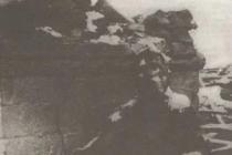
Computed tomograms in the upper mediastinum on the right revealed an additional soft tissue formation of irregular shape, with clear contours, extending to the neck area with a cystic component in its structure, with a total size of 30x14x23 mm (V=5 cm 3). The formation moderately evenly accumulated a contrast agent from 55-60 HU in the native phase of scanning to 90 HU in the arterial, and up to 80 HU in the venous phase.
Thymus in a typical place was not determined.
The identified changes in the mediastinal area according to MSCT data must be differentiated between a neoplasm of the upper mediastinum and thymus ectopia.
The child underwent surgery: Removal of a neoplasm of the upper mediastinum. According to the results of the histological conclusion of the remote formation: normal thymus tissue.
List of used literature
- Nishino M, Ashiku SK, Kocher ON et al. The thymus: a comprehensive review. 2006;26(2):335-48.
- Moore AV, Korobkin M, Olanow W et al. Age-related changes in the thymus gland: CT-pathologic correlation. AJR Am J Roentgenol. 1983;141(2):241-6.
- Baron RL, Lee JK, Sagel SS et al. Computed tomography of the normal thymus. 1982;142(1):121-5.
- Popa GA, Preda EM, Scheau C et al. Updates in MRI characterization of the thymus in myasthenic patients. J Med Life. 2014;5(2):206-10.
- Ackman JB, Wu CC. MRI of the thymus. AJR Am J Roentgenol. 2011;197(1): W15-20.
- Simanovsky N, Hiller N, Loubashevsky N et al. Normal CT characteristics of the thymus in adults. Eur J Radiol. 2012;81(11):3581-6.
ORIGINAL ARTICLE
DOI: 10.15690/onco.v2i2.1341
T.R. Panferova, A.L. Nikulina, I.N. Serebryakova, V.G. Polyakov
Research Institute of Pediatric Oncology and Hematology Federal State Budgetary Scientific Institution “N.N. N.N. Blokhin, Moscow, the Russian Federation
Ultrasound diagnosis of ectopic thymus tissue in the thyroid
gland in children
For the study, 22 patients aged 1.5-16 years with sonographic signs of foci of ectopic thymus tissue in the thyroid gland were selected, which were observed at the institute in 2012-2015. In all children, the thymus gland was located typically, had a normal size and a normal structure, which corresponded to the structure of ectopic areas in the thyroid gland. The foci of thymic tissue ectopia in the thyroid gland were oval (89.7%) or irregularly shaped (10.3%) with clear (100%) uneven (79.3%) or even (20.7%) contours, low echogenicity. (100%), with uniform thin linear inclusions (100%) in combination with hyperechoic punctate inclusions (31.0%), avascular (86.4%) or moderately vascularized (13.6%). In 5 children with lesions larger than 10 mm, the diagnosis was confirmed by cytological examination after targeted fine-needle aspiration biopsy. The follow-up period was 9-36 months. In one child aged 7 years with an area of ectopia, confirmed by cytology, for 9 months of observation, a decrease in the volume of the focus by 34% was noted, in the remaining children no dynamics were detected. We believe that when identifying characteristic sonographic signs, it is legitimate to make an ultrasound conclusion about the ectopia of the thymus tissue in the thyroid gland. Ectopic foci larger than 10 mm require morphological verification. All children should be dynamically monitored to assess expected regression or rule out possible transformation.
Key words: thymus ectopia, thyroid gland, thymus gland, ultrasound diagnostics, children.
(For citation: Panferova T.R., Nikulina A.L., Serebryakova I.N., Polyakov V.G. Ultrasound diagnosis of ectopic thymus tissue in the thyroid gland in children. Oncopediatrics. 2015; 2 (2): 109-114 .doi: 10.15690/onco.v2i2.1341)
T.R. Panferova, A.L. Nilulina, I.N. Serebryakova, V.G. Polyakov
Institute of Pediatric Oncology and Hematology N.N. Blokhin, Moscow, Russian Federation
The Ultrasound Diagnosis of Ectopic Thymus Tissue in the Thyroid Gland in Children
Present study included 22 participants range in age from 1.5 to 16 years old with ultrasound features of an ectopic thymus in thyroid gland. The lesion structure was typical for the thymus in each case. The location and size of the thymus was normal. The ectopic lesions of thymus in thyroid gland had oval (89.7%) or irregular (10.3%) shape with sharp (100%) and irregular (79.3%) or regular (20.7%) contour, low echogenecity (100%), thin regular linear (100%) in combination with granular hyperechoic (31%) inclusions, avascularity (86.4%) or hypovascularity (13.6%). In 5 lesions larger than 10 mm the diagnosis was confirmed by fine-needle aspiration biopsy. The follow-up period was 9-36 months. In one case (the 7-year old boy) with confirmed diagnosis the lesion had been reduced on 34% during the follow-up period of 9 months. There were no changes in other patients during the follow-up. We consider it is reasonable to suggest the ectopic thymus in the thyroid gland if these ultrasound features are revealed. The lesions of ectopy more than 10 mm require the morphological confirmation. The long follow-up of these patients for expected regression or potential transformation is necessary. Key words: thymus ectopy, thyroid gland, thymus, ultrasonography, children.
(For citation: Panferova TR, Nilulina AL, Serebryakova IN, Polyakov VG The Ultrasound Diagnosis of Ectopic Thymus Tissue in the Thyroid Gland in Children. Onkopediatria. 2015; 2 (2): 109-114. Doi: 10.15690/onco.v2i2. 1341)
ORIGINAL ARTICLES
INTRODUCTION
Due to the widespread introduction in Russia of preventive ultrasound examinations (ultrasound) of the thyroid gland, conducted in children preschool age and adolescents, as well as due to the improvement in the quality of ultrasound equipment, the number of detected asymptomatic focal changes in the thyroid gland increased. In addition to diseases requiring surgical and / or medical treatment, such as cancer, adenoma, nodular goiter, chronic autoimmune thyroiditis with nodulation, the ultrasound diagnosis of which is well known and quite fully covered in the manuals, such an anomaly of development as ectopia of thymus tissue has become more often detected. -kovy gland. This phenomenon is well known to morphologists and embryologists, however, it often causes difficulties for ultrasound diagnostic doctors. Sonographic signs of ectopia of the thymus tissue into the thyroid gland have not found detailed coverage in the domestic scientific literature - there are only reports of individual cases, in foreign literature this topic is also not covered in sufficient detail.
The thymus gland, a lymphoid and endocrine organ that plays an important role in the formation of immunity, consists of two asymmetric lobes flattened in the anteroposterior direction, located in the anterior superior mediastinum. The left lobe is in most cases larger than the right lobe.
The thymus gland is formed by two germ layers - ectoderm and endoderm - from III and, to a lesser extent, IV pair of gill pockets at the 6th week of intrauterine development. The rudiments grow caudally and on the 8th week are displaced behind the sternum. With the persistence of the cranial part of the bud, additional (aberrant) thymus lobules can form, which are determined in the thickness of the thyroid gland, soft tissues neck, mediastinum. This anomaly is more common in the left side of the neck and mediastinum in females.
By the time of birth in full-term newborns, the thymus is formed and is represented by lobules inhabited by lymphocytes with well-defined cortical and medulla layers. The age-related involution of the thymus begins at the age of 5-8 years and ends by the puberty: the adipose tissue that appears in the gland gradually grows into the thymus lobules.
Information about the frequency of additional ectopic thymus lobules is contradictory. For example, Avula et al. report 9 cases of ectopic thymus detection among 287 children, which is 3.14%. According to Bale et al., this frequency is only 0.03% (one case in 3236 autopsies). According to H.G. Kim et al., this anomaly was found in 0.4% of children. Our own observations do not allow us to determine the frequency of thymus ectopia in the pediatric population, since in our medical institution
After identifying focal changes in the thyroid gland, patients are referred for a clarifying examination and deciding whether it is necessary to conduct a targeted fine-needle aspiration biopsy.
Foci of thymus ectopia are usually interpreted by ultrasound doctors as nodular hyperplasia, which sometimes leads to unreasonable prescribing of hormone therapy. Cases of cytological verification of areas of thymus ectopia without a correct assessment by an ultrasound doctor are rare, since the lymphoid elements detected in the punctate make it possible to erroneously assume autoimmune thyroiditis or lymphoma with thyroid lesions, while it is the combination of epithelial cells and lymphoid elements at various stages of differentiation. -tsirovki is proof of the thymic origin of the focus.
Histologically confirmed cases of thymus ectopia are presented in the literature usually as concomitant with the underlying disease for which surgery was performed: for example, hemithyroidectomy due to Carney syndrome (Carney complex). In these cases, sonography showed areas of ectopy similar to the tissue of the thymus gland - low echogenicity, uniform linear striation, dotted hyperechoic inclusions.
PATIENTS AND METHODS
Study participants
The study included children sent to our institute in 2012-2014. to clarify the nature of focal changes in the thyroid gland, identified during preventive examinations in other medical institutions. The referral institution diagnoses were "thyroid nodule" or "nodular goiter".
Research methods
For ultrasound of the thyroid gland, we used the generally accepted method of scanning and assessing the volume of the organ using ultrasonic scanners Phlips iU 22, Philips HD11XE, Siemens Acuson S2000.
For visualization of the thymus, combined scanning was used - from the retrosternal and parasternal approaches, for which microconvex and linear sensors were used with a frequency of 5-10 and 5-18 MHz, respectively.
For better visualization of the thymus from the retrosternal access, the child was placed on his back, placed under upper part chest low roller. Panoramic scanning was performed with a convex probe from the jugular notch: it made it possible to assess the transverse and anteroposterior dimensions of the thymus gland. To assess the size of the thymus, we used the standards proposed by T. Amour et al. and recommended by I.V. Dvoryakovsky. The transverse dimensions of the thymus are normally:
At 1-5 years - 33-57 mm;
6-10 years - 22-62 mm;
11-15 years - 34-54 mm;
16-19 years - 23-55 mm.
Visualization of the lower parts of the thymus lobes was performed by parasternal scanning with a microconvex probe from the intercostal spaces. The structure of the organ was assessed using ultrasound with linear probes from the retrosternal and parasternal approaches. Targeted fine needle aspiration biopsy (PTAB) was performed in 6 children. All children underwent control ultrasound examinations, the average follow-up time was 14.5 months.
RESULTS
Based on the fact that the ectopic thymus tissue has similar echo-signs with the normal thymus, we have developed preliminary criteria for the ultrasound diagnosis of thymus displacement into the thyroid gland: the presence of one or more focal asymptomatic oval formations similar in structure to the thymus gland; low echogenicity of the foci; uniform linear and/or granular inclusions; clear uneven or even contours; absence of liquid inclusions and perifocal changes.
We selected 22 children from 1.5 to 16 years old, in whom in the thyroid gland with ultrasound
The survey identified changes that meet these criteria. In 20 children, focal formations were found during preventive examinations, in 2 - during examinations for other diseases: one of them was sent for ultrasound of the thyroid gland due to obesity of the 1st degree, the other was examined for atopic dermatitis.
Characteristics of patients are presented in table.
The distribution of children by gender did not reveal a significant difference: 12 girls and 10 boys (54.5 and 45.5%, respectively).
On examination, none of the children had clinical signs of thyroid dysfunction. Volumetric formations during palpation of the thyroid gland were not determined. The level of hormones (TSH, T3, T4) and antibodies to thyroperoxidase and thyroglobulin in all patients was within normal limits.
The size of the thyroid gland in all patients was within the normal range, the structure of the gland was homogeneous, of medium echogenicity; the degree of vascularization corresponded to age. The size, shape and structure of the thymus gland in all children were normal.
A total of 29 foci were found that had ultrasound signs of ectopia, while they were located bilaterally in 4 children. The dimensions of the long axis of ectopic foci ranged from 3 to 25 mm (mean 8.2 mm, median 7 mm).
Table. Characteristics of patients with thymus ectopia in the thyroid gland
No. Age, years Sex Time of follow-up, months Indications for ultrasound of the thyroid tissue Localization/Dimensions
Left lobe, mm Right lobe, mm
1 6 F and Medical examination 10x3x4 -
2 1.5 M 12 Clinical examination 7x5x5 -
3 7 F 14 Medical examination 6x3x3 -
4 6 F 12 Medical examination 3x3x1 3x3x2
5 13 F 12 Clinical examination - 6x4x4
6 5 F 14 Medical examination 8x3x5 and 3x2x3 -
7 14 F 14 Clinical examination 11x7x9 -
8 13 М 14 Obesity I degree 9x3x7 -
9 6 M 13 Clinical examination 13x6x9 -
10 7 F 11 Clinical examination 3x2x3 3x1x3
11 13 F 15 Clinical examination 15x7x10 -
12 7 М 9 Medical examination 4x2x2 25x8x9
13 6 F 13 Clinical examination 8x5x6 -
14 8 M 9 Medical examination 7x2x5 and 8x3x5 -
15 6 F 13 Clinical examination 7x3x3 -
16 4 М 36 Clinical examination - 6x3x6
17 16 F 9 Clinical examination - 10x4x6
18 7 М 13 Clinical examination 11x7x7 -
19 4 M 29 Atopic dermatitis - 6x4x6
20 6 M 17 Clinical examination 4x2x3 6x3x4
21 6 F 10 Clinical examination 20x4x10 -
22 7 M 19 Medical examination 8x6x7 and 7x6x7 -
ORIGINAL ARTICLES
Rice. 1. Patient, 7 years old. The similarity of the structure of the thymus (A - transverse sonogram from the retrosternal access) and the site of ectopia in the left lobe of the thyroid gland (B - longitudinal sonogram)
In 6 cases (20.7%), large foci (> 11 mm along the long axis) were detected, in 23 (79.3%) - small (< 10 мм). В левой доле выявлен 21 (72,4%) участок эктопии, в правой - 8 (27,6%). В 16 (55,2%) случаях очаги эктопии располагались в средних отделах долей, в 10 (34,5%) - прилежали к заднему контуру железы, в 3 (10,3%) - выходили на контур в области полюсов долей (2 - к верхним полюсам левой и правой долей, 1 - к нижнему полюсу правой доли).
PTAB was performed in all children with ectopic areas exceeding 10 mm along the long axis (6 patients). In 1 child, the puncture was uninformative, in 5 children a mixed-cellular composition was found in the punctate, represented by epithelial cells and lymphoid elements at different stages of differentiation, which made it possible to confirm the sonographic diagnosis of thymic tissue ectopia in the thyroid gland. Patients with small lesions underwent only follow-up ultrasound.
Since the selection of focal formations followed the criteria described above, in all cases (100%) there was a correspondence between the structure of the thymus and focal formations in the thyroid gland: low echogenicity; thin regular linear inclusions forming a uniform mesh pattern; clear contours; absence of perifocal changes in the thyroid gland, such as compression of the surrounding parenchyma, hypoechoic rim, increased vascularization, etc. (Fig. 1).
In most cases (23; 79.4%), the shape of the identified foci was oval, flattened in the anteroposterior direction (Fig. 2). An irregular shape (Fig. 3) or a regular oval shape were less common - 3 cases each (10.3%). Large foci (> 10 mm along the long axis; Fig. 4, 5) had an oval regular shape.
Uneven, partially pointed contours were detected in small lesions (23; 79.3%), even in large areas of ectopia (6; 20.7%). Along with linear inclusions, which were determined in all cases, in 9 foci of ectopia
(31.0%) had pinpoint hyperechoic inclusions. It should be noted that the structure of ectopic foci fully corresponded to the structure of the thymus gland in each individual child.
In 25 (86.4%) areas of ectopia, blood flow was not recorded by color Doppler mapping and power Dopplerography, in
Rice. 2. Patient, 6 years old. Longitudinal sonogram of the left lobe of the thyroid gland. Small oval flattened ectopia focus (6x3x4 mm)
Rice. 3. Patient, 14 years old. Longitudinal sonogram of the left lobe of the thyroid gland. Irregularly shaped ectopic focus with uneven pointed contours, dimensions 11x7x9 mm
Rice. 4. Patient, 7 years old. Longitudinal sonogram of the right lobe of the thyroid gland. Large ectopic area (25x8x9 mm), oval in shape with smooth contours, located in the upper pole of the lobe
ONCOPEDIATRY / 2015 / volume 2 / № 2
Rice. 5. The same patient. Transverse sonogram at the level of the upper pole of the right lobe of the thyroid gland
Rice. 6. The same patient. Transverse sonogram with color Doppler mapping of the right lobe of the thyroid gland. In the thickness of the ectopic thymus tissue, a moderate number of small vessels is determined.
Rice. 7. Patient, 6 years old. Papillary thyroid cancer.
In the lower parts of the right lobe and the isthmus, a tumor of low heterogeneous echogenicity with uneven and indistinct contours and multiple dotted microcalcifications is determined
In 4 (13.6%) large areas, single small vessels were observed (Fig. 6).
All children underwent repeated clinical examinations and ultrasound of the thyroid gland. During the observation period, the size of 28 foci (96.6%) of thymus ectopia did not change. One 7-year-old child with a large ectopic focus (25x8x9 mm) confirmed morphologically had a 34% reduction in the mass during 9 months of follow-up.
Rice. 8. The same patient. Metastatic lesion of the upper jugular lymph nodes of the contralateral side
DISCUSSION
Thyroid nodules are rare in children. The frequency of their detection in the children's population is 0.2-5.1%, in contrast to the adult population, where they occur in 40-65% of cases. However, thyroid cancer is more common in children than in adults (22% versus 14%). If the information of Avula et al. about the frequency of occurrence of an ectopic thymus, given at the beginning of our article (3.14%), will be the closest to reality, then this anomaly can take first place in the frequency of detection in the thyroid gland in children.
We believe that sonographic identification of thymic ectopic foci in the thyroid gland in children is of great practical importance.
If small foci are detected in the thyroid gland that have all the typical ultrasound signs of ectopia, it is possible to avoid an invasive procedure under anesthesia for children (PTAB) and limit yourself to follow-up ultrasound examinations once every 3 months for six months, and then once a year.
The correct interpretation of the nature of these foci will allow avoiding the diagnosis of "Nodular goiter" on duty in such cases, which often causes unnecessary anxiety in patients and their parents, and sometimes unreasonable drug treatment.
If large areas (> 10 mm) are identified, PTAB is recommended to rule out thyroid cancer and confirm thymic ectopia. It is known that papillary thyroid cancer may have a similar picture with ectopic foci, namely: low echogenicity, linear and point hyperechoic inclusions, irregular shape, uneven contours. Cases are described in the literature when a cytological examination of formations doubtful in relation to cancer confirmed ectopic thymus tissue. Distinctive signs of cancer from ectopia are heterogeneity, fuzzy contours, micro-calcifications in the structure of the tumor, and possible metastatic lesions of the lymph nodes of the neck (Fig. 7, 8).
ORIGINAL ARTICLES
We consider the sonographic diagnosis of thymus ectopia in the examined children to be convincing if the following criteria are present: identification of flattened oval (79.4%), regular oval (10.3%) or irregularly shaped (10.3%) foci in the thyroid gland with clear (100 %) uneven (79.3%) or even (20.7%) contours, low echogenicity (100%) with hyperechoic multiple thin linear inclusions (100%), forming a fine mesh pattern, in combination (31.0%) with hyperechoic punctate inclusions, avascular (86.4%) or moderately vascularized (13.6%). Ectopic foci correspond to the structure of the thymus in each individual patient, do not cause a volume effect on the surrounding parenchyma of the thyroid gland.
noah gland, do not have liquid zones. In the presence of the described picture and foci less than 10 mm along the long axis, dynamic ultrasound control can be limited. Lesions larger than 11 mm should be subjected to PTAB to rule out thyroid cancer.
In the future, we propose a long-term follow-up of patients with foci of thymus ectopia in the thyroid gland to study the natural development of this condition, assess the expected regression, or identify possible tumor transformation.
LITERATURE
1. Pykov M.I., Vatolin V.K. Children's ultrasound diagnostics. M.: Vidar. 2001. S. 557-561.
2. Kharchenko V.P., Kotlyarov P.M., Zubarev A.R. Diagnosis of thyroid cancer by ultrasound. Moscow. 2002, p. 72.
3. Tovi F., Mares A.J. The aberrant cervical thymus: embryology, pathology, and clinical implications. Am J Surg. 1978; 136:631-637.
4. Shcherbinina V.I., Stepanova E.A., Banina V.B. and others. Cervical localization of the aberrant thymus gland. Children's surgery. 2009; 1:52-52.
5. Kim H.G., Kim M.J., Lee M.J. et al. Sonographic appearance of intrathyroid ectopic thymus in children. J Clin Ultrasound. 2012; 40(5):266-271.
6. Bale P.M., Sotelo-Avila C. Maldescent of the thymus: 34 necropsy and 10 surgical cases, including 7 thymuses medial to the mandible. Pediatric Pathol. 1993; 13:181.
7. Avula S., Daneman A., Navarro O.M. et al. Incidental thyroid abnormalities identified on neck US for non-thyroid disorders. Pediatric Radiol. 2010; 40:1774.
8. Park S.H., Ryu C.W., Kim G.Y. Intrathyroidal thymic tissue mimicking a malignant thyroid nodule in a 4-year-old child. ultrasonography. 2014; 33(1):71-73.
9. Megremis S., Stiakaki E., Tritou I. et al. Ectopic intrathyroidal thymus misdiagnosed as a thyroid nodule: sonographic appearance. J Clin Ultrasound. 2008; 36:443-447.
10. Courcoutsakis N., Patronas N., Armando C.F. et al. Ectopic Thymus Presenting as a Thyroid Nodule in a Patient with the Carney Complex. thyroid. 2009; 19(3):293-296.
Sweaty feet! Horror! What to do? And the way out is very simple. All recipes that we give are tested first of all on ourselves and have a 100% guarantee of effectiveness. So, get rid of sweaty feet.
There is much more useful information in the history of the patient's life than in all the encyclopedias of the world. People need your experience - "the son of difficult mistakes." I ask everyone to send prescriptions, do not spare advice, they are a ray of light for the patient!
ABOUT medicinal property pumpkin Ingrown toenail I am 73 years old. Sores appear such that I did not even know that they existed. For example, on the big toe, a nail suddenly began to grow. The pain kept me from walking. They suggested surgery. In "Healthy Lifestyle" I read about pumpkin ointment. I cleaned the pulp from the seeds, applied it to the nail and bandaged it with polyethylene so that the […]
Fungus on the legs Fungus on the legs Pour hot water into the basin (the hotter the better) and rub laundry soap in the water with a washcloth. Hold your legs in it for 10-15 minutes to steam them properly. Then clean the soles and heels with a pumice stone, be sure to trim your nails. Wipe your feet dry, dry and lubricate them with a nourishing cream. Now take a pharmacy birch […]
For 15 years, my foot has not bothered me. Callus on my leg For a long time, I was bothered by a callus on my left foot. I cured him in 7 nights, got rid of the pain and began to walk normally. It is necessary to grate a piece of black radish, put the gruel on a rag, firmly tie it to a sore spot, wrap it with cellophane and put on a sock. Compress is desirable to do at night. To me […]
Young Doctor Prescribed His Grandmother's Prescription Gout, Heel Spurs I'm sending you a prescription for a heel spur and lumps near the big toe. It was given to me by a young doctor about 15 years ago. He said: “I can’t write out a sick leave certificate for this, it’s not supposed to. But my grandmother was treated for these troubles in such a way ... ”I took the advice […]
Let's start with gout, which is caused mainly by a violation of metabolic processes. Let's listen to what the Vinnitsa doctor D.V. NAUMOV says about padagra. We treat gout according to Naumov Gout "healthy lifestyle": There are a lot of questions about the dissolution of salts in the joints. You claim that the food salt that we use inside has nothing to do with insoluble salts such as urates, phosphates and oxalates. And what has […]
On the advice of Antonina Khlobystina Osteomyelitis At the age of 12, I fell ill with osteomyelitis and almost lost my leg. I was admitted to the hospital in a serious condition and operated on the same day. He was treated for a whole month, and was deregistered only after 12 years. I was cured after all by a simple folk remedy, which was suggested to me by Antonina Khlobystyna from Chelyabinsk-70 (now […]
Fell, woke up - gypsum Over the years, the bones become very fragile, osteoporosis develops - women especially suffer from this. What to do if you have a fracture? How can you help yourself besides plaster and bed rest? With these questions, we turned to Doctor of Biological Sciences, Professor Dmitry Dmitrievich SUMAROKOV, a specialist in bone tissue restoration. "ZOZH": You are 25 years […]
Onion soup against osteoporosis Osteoporosis Doctors call osteoporosis the "silent thief". Quietly and without pain, calcium leaves the bones. A person has osteoporosis and knows nothing about it! And then unexpected bone fractures begin. A 74-year-old man was admitted to our hospital with a hip fracture. He fell in an apartment out of the blue - the bone could not withstand the […]
Dear visitors of the portal! In the archive of medical consultations for 13 years, there are a large number of prepared materials that you can use. best regards, editorial
The section "consultations" suspends its work.
Eugene asks:
Hello. The boy is 7 years old. Today they did an ultrasound, they said that the shield of the gland was increased to 12 cm, at a rate of 4. I have no more data. Passed a blood test for TSH, T4 St and ATkTPO. Waiting for a response. (The husband left the ultrasound with the doctor). Dz was previously written like this: endemic goiter 1st. and Chr AIT? Acceptance after analysis. How serious is it that the shield of iron is so greatly increased? The child is normal and develops normally. I don't see any deviations. Tell me, is this serious?
Answers:
Hello Evgenia! In fact, at the moment we have nothing to base our assumptions on. Even the result of ultrasound you write from the words of your husband, and the figure of 12 cm raises serious doubts. Therefore, at this stage, we can not say anything specific. AIT (autoimmune thyroiditis) and endemic goiter are treatable diseases. Therefore, you just need to wait until the end of the examination and start treating the child. Take care of your health!
Julia asks:
Ultrasound examination of the thyroid gland: usually located, left lobe length 33.2 width 9.6 thickness 10.7 volume 1.6 right lobe length 37 width 10.7 thickness 11.4 volume 2.2 isthmus 2.7 echogenicity is normal, lymph nodes are not enlarged. A fiery cyst with a diameter of 5.9-3.1 was found c / z of the left lobe of the bronchi; in conclusion, a non-thyroidal formation on the left. Blood test TSH-1.15. What does this mean, what are our further actions with the child? A year ago, during an ultrasound of the thyroid done by a thyroidologist, a thyroid ectopia of the thymus was detected outside the thyroid formation, the doctor then told me that this was a feature of nothing terrible, he did not see, and today it was not a thyroidologist who did the ultrasound, the endocrinologist advised us to consult with local surgeons and take iodomarin daily, see the endocrinologist in six months . Tell me what to do with the cyst, what are our next steps?
Responsible Berezhnaya Irina Yurievna:
Hello Julia First of all, it is necessary to clarify the diagnosis: thymus (hardly ectopia, most likely visualization) or ectopic tissue of the thyroid gland. More trustworthy is the conclusion of a doctor a year ago, since there is no bronchiogenic cyst in the thyroid gland. You just need to do a CT scan (MRI) of the neck area and consult with a pediatric endocrinologist. I hope that the surgeon's help here will only be needed advisory. Just investigate.
asks Elena, 31 years old, Saratov:
Hello, please tell me! My daughter is 6 years old, height 129 cm, weight 32 kg. They tested for hormones, as a result of TSH-6.85 mIU / l, T4-20.8 nmol / l.
tell me, I beg you, the increased TSH may be due to the fact that the height and weight of my daughter is much greater than that of her peers?
Does the discrepancy between the height and weight of the child for age affect the level of the hormone TSH?
Answers:
Hello, Elena. The TSH hormone regulates the functioning of the thyroid gland, in this case it is elevated (there is a reduced work of the thyroid gland), which can be manifested by a decrease in the rate of metabolic processes (including weight gain). It is necessary to do an ultrasound of the thyroid gland and antibodies (analysis of ATPO, AMS, ATTG), growth hormone STH and, if necessary, an x-ray of the hands to determine the compliance of the bone age with the passport one. Sincerely, Natalya Vasilievna.
Gulmira asks:
Hello. I am 32 years old. In 2011, I was diagnosed with primary hypothyroidism. I am currently taking Euthyrox 50mg. In my daughter's analysis: TSH - 14.7 μIU / ml, T4 free - 12.1 Pmol / l what does this mean? what drugs should we take? My daughter is growing according to her age.
Responsible Medical consultant of the health-ua.org portal:
Hello, the results of your child's tests indicate the presence of subclinical hypothyroidism (high TSH). It is necessary to contact a pediatric endocrinologist for further additional examination and therapy. Children with subclinical hypothyroidism are also shown to prescribe replacement therapy thyroid hormones and iodine preparations, after establishing the cause. The appointment of therapy is carried out only by a pediatric endocrinologist with careful selection and adjustment of doses.
Vladimir asks:
Good afternoon, tell me - is it worth giving a 3.5-year-old child iodamarin for prevention ?? and if so, how much?
Responsible Medical consultant of the health-ua.org portal:
Hello Vladimir! If you live in an area with insufficient iodine in the environment, iodine prophylaxis will not hurt. Children under 12 years of age can be given iodomarin at a dose of 50-100 mg per day, after agreeing the dose of the drug with the attending physician (pediatrician). Perhaps your child is already receiving a multivitamin-mineral complex, which includes iodine, in which case the dose of iodine should be adjusted taking into account the iodine content of this preparation. Take care of your health!
Christina asks:
Good afternoon! Daughter 6 years 7 months. Height 121, weight 22. Bone age 7 years. They did an ultrasound of the thyroid gland. Dimensions: right lobe 0.68 cm, thickness 0.76 cm, length 2.26 cm, volume 1.2 cm in 3. Left lobe width 1.03 cm, thickness 0.66 cm, length 2 .05 cm, volume 1.7 cm in 3. Isthmus thickness 0.13 mm, total volume 1.4 ml. We were referred to an endocrinologist. Is there any deviation from the norm. Thank you!!!
Responsible Berezhnaya Irina Yurievna:
Hello Christina The total volume of an organ in a child aged 6-14 should be 4.9-5.0 cm3 (Brunno). When measuring a volume that differs from statistical data, an endocrinologist's consultation is certainly needed. The purpose of the consultation: whether these data are physiological characteristics or additional examination is necessary (presence of iodine in the urine, TSH level in the blood ...). It is not excluded repeated ultrasound examination of the thyroid gland in a specialized center.
Tatyana asks:
Hello! Please help me figure it out. Ultrasound showed that vascularization is enhanced during color doppling. right lobe-v=1.8cm3, left lobe v=1.6cm3. handed over for hormones TSH = 1.6 mMe / l,
T4= 22.0pmol/l, T3=7.2pmol/l, ATPO=0.
The child has reduced weight, constant hand tremor, heart palpitations. In the clinic, the endocrinologist said that the child is fine, and the hand tremor is age-related. Help with advice.
Responsible Berezhnaya Irina Yurievna:
Hello, Tatyana Examination of the thyroid gland revealed no abnormalities: ultrasound and hormonal examinations are unchanged. The endocrinologist's task is to exclude thyrotoxicosis and pheochromocytoma (tremor can be detected in these diseases). Unfortunately, you did not indicate the age of the child. In any case, you need the help of a pediatric neurologist. tremor can be both a consequence of the immaturity of the nervous system (physiologically) and early signs of disease.
Hope asks:
Hello.
The child was born at 41-42 weeks 3 kg 360. Tight entanglement of the umbilical cord. Prolonged jaundice 325. Treatment for 3 weeks, discharge at 140.
Analyzes and ultrasound May 2014. Daughter 8.5 years old 29kg 138cm
TSH 6.36 T4sv 10.17 a/t to TPO 0.3 TG 0.2.
Ultrasound of the thyroid gland (28.05.14) Increased in size, diffuse cystic changes in the parenchyma. Total volume 5.36
Treatment: Vit B1, V6v / m No. 10, Elkar, Pantogam 0.25 3 r per day, Iodomarin 3 months, glycine.
Repeated tests November 2014. Daughter 142cm 32kg 9 years
ST4 13.0 TSH 6.9
Previously, the daughter was healthy, for 2 years now she has been feeling unwell, complaints: weakness, headaches, sweating, pain in the abdomen and heart, soreness and discomfort in the throat.
They also did an ultrasound of the abdominal cavity (03/25/14)
Conclusion: Signs of cholecystitis cholongitis. Signs of superficial gastritis. Reactive changes in the pancreas. (Addition: Severe flatulence)
Please help me to decipher and understand our results, analyzes, and can it be that the problems of the abdominal cavity that we are concerned about are related to thyroid problems? I am also very worried about perspiration, suffocation, discomfort in the throat.
Thank you very much in advance for your advice.
Responsible Berezhnaya Irina Yurievna:
Hello Nadezhda In my opinion, the child can be diagnosed with subclinical hypothyroidism (SH), which requires the appointment of L-thyroxine. All accompanying complaints can be associated with SH: a decrease in the secreto- and acid-forming function of the stomach and a significant increase in the content of mucoproteins and a decrease in the ratio of phospholipids/cholesterol in serum. The prescribed treatment is inadequate. A specialized consultation with an endocrinologist is required.
Inna asks:
Hello, please decipher the tests! How scary is everything? and what to do? The child is 7 years old, height 115, weight 23 kg. FT4-16.32 and TSH-5.16 Many thanks in advance!
Responsible Medical consultant of the health-ua.org portal:
Hello Inna! It is not possible to interpret the results of the above study, because you did not indicate the sex of the child, did not provide units of measurement and data on the reference (normal) limits used in the laboratory that performed the analysis (printed on the analysis form to the right or left of the study results). Complete your message with the necessary information or discuss the results of the analysis with your doctor. Take care of your health!
Elena asks:
Hello! My girl is 10 years old. According to the measurements of the endocrinologist, she looks 12.5 years old (height - 152 cm, weight - 68 kg). They took a blood test for hormones. T4 free - 9.17 pmol / l, TSH - 4.43 μU / ml and we were diagnosed with exogenous constitutional obesity E66.0: 2 degrees and subclinical hypothyroidism E02.0
The doctor prescribed us Euthyrox 75 mcg.
Prompt please - whether it is necessary to us at such results to sit down on hormones.
Responsible Renchkovskaya Natalya Vasilievna:
Hello, Elena. The girl needs to do an ultrasound, in the tests it is necessary to exclude autoimmune thyroiditis (an for antibodies AMS, ATPO), the dose of euthyrox is prescribed with a minimum (half a tablet from a dose of 25) or a homeopathic option so as to balance the level of TSH. It is also important to assess the appropriate bone and passport age, the degree of sexual development and write down an individual balanced diet. With uv. Natalya Vasilievna.
Zlata asks:
Good afternoon. The child is 1 year and 7 months old, tested for thyroid hormones: T3- 1.72; T4 - 10.22; TSH- 1.63; ATTG-1034.98; ATTPO- 720.08; Ultrasound of the thyroid gland: Isthmus - 1.06
Right lobe (length 21.4; width 8.41; thickness 10.2; U of the right lobe 0.88; not enlarged). Left lobe (length 19.6; width 9.54; thickness 10.6; U of the right lobe 0.95; not enlarged). The total U thyroid 1.8 is not increased. It is typically located, the contours are clear, even. The capsule of the thyroid gland is not thickened, not compacted. the structure is homogeneous, echogenicity is normal. What can cause such blood tests and what consequences can this lead to? Can eat ways to normalize antibodies?
Responsible Volobaeva Ludmila Yurievna:
Good health! It is important to understand the purpose for which antibodies were determined for such a small child, whether relatives have thyroid diseases. An increase in antibodies is only a risk factor for the development of hypothyroidism in the future, but there is no need to reduce them. Medicine, at this stage of development, is not capable of doing this. In such a situation, it is only necessary to control TSH once every 6 months and ultrasound of the thyroid gland once a year. Antibodies do not need to be treated.
Oleg asks:
Hello! Girl 6 years old, height 122.5 cm, weight 30 kg. Ultrasound showed an increase in the thyroid gland by 34% + cholesterol readings were overestimated by 5.28 (mmol / l). On the basis of the analyzes, a course of iodomarin was prescribed by the doctor in the polyclinic. It would be desirable to receive your references, leaning against the given results of analyses. Regards, Oleg.
Responsible Kravchun Nonna Alexandrovna:
Hello Oleg! The reasons for the enlargement of the thyroid gland can be different - inflammation, autoimmune process, lack of iodine. There is not enough information to make a diagnosis - you need to study the function of the thyroid gland - donate blood for T3, T4, TSH, ATPO and AT-TG (thyroid profile). Only in combination with the results of these tests will it be possible to establish the true cause of the enlargement of the gland and prescribe the appropriate treatment. Do not be ill!
Ultrasound examination of the thyroid gland: when and why to do it
The thyroid gland is without a doubt one of the most important organs in the human body. The production of hormones responsible for metabolism and energy depends on its functioning. If something is wrong with the thyroid gland, then the consequences will manifest themselves in the most unexpected areas of the body's life.
To find out the cause of the failure in the gland, you need to contact an endocrinologist. The doctor will prescribe tests and an ultrasound of the thyroid gland.
Why do you need to do an ultrasound of the thyroid gland
A fairly common pathology of the thyroid gland is hypothyroidism, provoked by iodine deficiency. When this element enters the body not regularly, but with malfunctions, the first signs are not long in coming: hair falls out, skin dries, nails break, swelling and problems with sleep appear.
Suspect thyroid problems if some of the following symptoms are found:
- apathy;
- lethargy;
- mood swings;
- weakness;
- drowsiness;
- cardiac arrhythmia (see Thyroid gland and heart - what is the connection?);
- changes in weight (if there are not enough hormones, then excess weight appears, and when elevated level hormones are depleted).
The spectrum of manifestations of other pathologies of the thyroid gland may include many other symptoms.
In case of suspected thyroid disease, an ultrasound will be one of the first tests that a doctor will prescribe. Ultrasound examination is the study of any organ without penetration through the skin or mucous membranes of a person using ultrasonic waves.
Ultrasound examination will help to find out parameters such as:
- The structure of the thyroid gland. Normally, the organ consists of two lobes and the isthmus connecting them. Often there is a pyramidal part (lobe), located next to the gland or just above the isthmus. Perhaps the presence of tissue outgrowths, about a centimeter long, they are located near the lower edges of the lobes to the thymus gland. In prenatal development, a failure in the formation of the thyroid gland may occur, and as a result, there is no division into two lobes, this deviation is called aplasia (agenesis) of the lobe in the thyroid gland. If the development of the gland is absent, then the diagnosis is “complete aplasia”.
- The position of the thyroid gland is divided into typical, low, pathological (aberrant). Ectopia is also possible - the tissues of the thyroid gland go beyond the boundaries of their organ, which is possible with both the lobes and the isthmus.
- Contours, normally, should be clearly visible. If they are fuzzy, then this may indicate inflammation or a tumor.
- The structure should be homogeneous, with its inherent graininess. Heterogeneity can be a signal of an inflammatory process.
- Dimensions of the thyroid and parathyroid glands.
- Echogenicity - tone and shade of thyroid tissue.
- The formations are examined in case of the presence of nodes, cysts (see Cyst in the thyroid gland: symptoms and signs of pathology), or calcifications.
- Particular attention is paid to the presence, size, structure and structure of lymph nodes accessible to the apparatus, which can reveal microcalcifications, increased blood flow and other signs of the process of tumor formation.
- The structure of the salivary gland located near the ears.
- The size and structure of the larynx, as well as the soft tissues of the neck - to study the areas around the thyroid gland.
About undergoing a thyroid ultrasound procedure
So, how do you prepare for an ultrasound examination of such a tiny organ as the thyroid gland? The answer is simple: no way. No special action is needed. However, it is better for both young children and the elderly to have an ultrasound scan on an empty stomach, otherwise the gag reflex can be uncomfortable.
For the procedure, you need to take a towel with you: first, you can make a roller out of it and put it under your neck for greater convenience, and then it’s convenient for them to wipe the gel from your neck. If this procedure is not the first for the subject, then the previous ultrasound results should also be taken with you.
The very procedure of what is happening is as follows: throwing back his head, the subject lies down on the couch. On the neck, the specialist applies a special gel designed for a better ultrasound signal.
Then the study itself is carried out. The signal of a special sensor that works with ultrasonic waves is reflected from the thyroid tissue and then read by the same sensor. The signal is then examined and the result appears on the screen as an image of the thyroid gland.
The process does not cause any painful sensation, however, there may be discomfort due to the uncomfortable position of the head and neck. The whole process takes approximately 30 minutes.
It is often possible to get a free ultrasound in city clinics, but those who do not want or cannot wait in line can go to a paid clinic. The price depends on the city. For preventive purposes, it is enough to do an ultrasound examination once a year.
Significance of the obtained results of ultrasound examination
In the presented photo of the ultrasound of the thyroid gland, you can see it in different projections. The dotted line marks the contours, and the arrows mark the size of the shares. The pictures "a" and "c" show the transverse and longitudinal sections of the thyroid gland, the photo "b" shows the asymmetry of the lobes of the gland, and the picture "d" shows the normal vascular pattern.
Of course, only a specialist should decipher the results of ultrasound, since there are many large vessels in the human neck, The lymph nodes, thyroid, parathyroid and salivary glands, as well as organs such as the trachea and esophagus. However, some parameters can be understood by knowing the norm in advance.
The structure of the thyroid gland should be even, homogeneous. For men, the normal volume of the thyroid gland, according to medical indicators, should not exceed 25, and for women, on average - But you need to understand that these are only average indicators that fluctuate depending on the weight of the subject.
The table below will help guide you:
For children under 16 years old, other sizes are characteristic. It must be borne in mind that the size of the thyroid gland in boys and girls of the same year of birth is different, the difference can reach 1.5.
Approximate norms for the volume of the gland in children aged 6 to 15 years are as follows:
The thickness and size of the lobes in the thyroid gland can also be of different sizes, but among them there are also average indicators:
- the approximate size of the shares is 4x2x2 cm;
- the right and left shares should also be equal to each other;
- the norm of the thickness of the isthmus is considered to be the size of 4-5 mm.
Also at healthy person, according to the results of ultrasound:
- the contours of the thyroid gland are even and clearly visible;
- cervical lymph nodes are normal;
- the parathyroid glands are about 4x5x5 mm in size.
You can learn more about the results of ultrasound of the thyroid gland from the video in this article.
Thyroid diseases that can be detected by ultrasound
When ultrasound diagnostics of thyroid gland diseases displays a deviation from the indicators correlated with the norm on the monitor, this may indicate such diseases of the endocrine system:
- Thyroiditis. An enlarged thyroid gland and swelling are more likely to indicate thyroiditis due to bacteria and viruses. Symptoms can also be fever and headaches, as well as pain in the thyroid gland.
- Hypothyroidism. A reduced size of the gland indicates hypothyroidism and a decrease in the efficiency of the thyroid gland. When taking medications prescribed by a doctor, the package of which contains instructions, you can make up for the lack of hormone production.
- Inflammation is characterized by echogenicity above the norm and heterogeneity of the structure of the gland.
- Cyst. A rounded cavity with fluid inside, and clear edges and a normal structure on the outside - this is a typical description of a cyst. To find out if there are cancer cells in the cyst, the doctor may prescribe a puncture, that is, a collection of contents.
- Cancer tumors. Formations with high density and jagged edges. Enlarged lymph nodes may also indicate tumor growth. The tumor can be either benign (adenoma) or malignant (cancer).
- Diffuse-toxic goiter. Increased volume of the gland, mood swings, nervousness, weight loss are indicators of goiter caused by increased production of iodine-containing hormones from the gland.
- Nodular goiter. It can be felt by ordinary palpation. The seal is easily felt in the front of the neck. In the ultrasound picture, the goiter can be seen as a high-density focus with clear boundaries and healthy tissue.
But it should be understood that an ultrasound examination of the thyroid gland, the decoding of which is multi-stage and complex, can only be correctly read by a specialist who can also prescribe blood tests for hormones, sugar levels, general analyzes blood and urine.
Problems with the thyroid gland in modern life are quite common. Ultrasound examination, at the moment, is recognized as the most painless and safest method for examining the gland, and its location guarantees simplicity and convenience.
Ultrasound helps to detect the presence of diseases and disorders of the thyroid gland, as well as deviations in size from the required norm. All this helps to maintain the health of yourself and your loved ones.
Feeling of suffocation and a lump in the throat with pathologies of the thyroid gland
An attack of suffocation in pathologies of the thyroid gland
The feeling of suffocation, the sensation of a lump in the throat in case of thyroid gland pathologies is determined by a set of symptoms and factors:
- an attack of excitement;
- active labor;
- exposure to chemicals:
- medicines;
- food additives.
The attack is accompanied by the following symptoms:
- pressure in the throat when swallowing;
- dryness, burning in the mouth;
- sore throat, coughing;
- easy suffocation with lack of air;
- there is weakness, sweat comes out.
Causes
To determine the cause of the feeling of a lump in the throat, it is necessary to exclude the pathologies of the organs that are accompanied by this symptom:
- 1. Quincke's edema. This manifestation of allergy often causes a feeling of a foreign body getting into the larynx, difficulty breathing. A history of rashes confirms a tendency to allergies. It is necessary to urgently take a large dose of an antihistamine drug, call an ambulance.
- 2. Feeling of a lump in the throat can cause vegetovascular dystonia. Attacks are provoked by nervous experiences, weather changes. This disease is confirmed by the presence of the following symptoms: nausea; dizziness; apathy. Attacks are provoked by nervous experiences, weather changes.
- 3. Discomfort in the form of a burning sensation may be heartburn caused by the reflux of gastric juice through the esophagus into the lower pharynx. Frequent heartburn confirms acidity and gastritis. It is possible to alleviate the condition in this case by reducing the amount of salty, sweet, fried foods consumed.
- 4. The state of lack of air accompanies attacks of diseases such as asthma, SARS, pharyngitis and causes discomfort in the larynx.
Thyroid disease
Diseases, accompanied by a feeling that the thyroid gland is choking, are divided by endocrinologists into three types:
- 1. Thyroiditis.
- 2. Nodular goiter.
- 3. Diffuse goiter.
diffuse goiter
Pathology is easily determined by visual examination due to a significant increase in the volume of the thyroid gland.
The organ is located close to the skin of the anterior region of the neck, so its growth is imperceptible only with excessive deposition of fat in the neck.
Diffuse goiter is accompanied by the following symptoms:
- discomfort while eating;
- bouts of dry cough;
- hoarseness of voice;
- discomfort if clothes press on the throat.
In this disease, the entire tissue of the organ is affected, which leads to a violation of the synthesis of hormones. A decrease in thyroid hormones in the venous blood leads to hypothyroidism, manifested by:
- weight gain;
- tissue swelling;
- shortness of breath;
- bradycardia;
- loss of eyelashes and eyebrows;
- pallor and dry skin.
An excess of thyroid hormones is accompanied by thyrotoxicosis, causing:
- weight loss;
- sleep disorders;
- tachycardia;
- skin moisture;
- anxiety;
- hand tremor.
nodular goiter
This pathology leads to partial damage to the tissues of the thyroid gland, causing the formation of nodes.
A node is an increase in the structural unit of an organ - a follicle.
By the number of nodular formations, this pathology is divided into types:
- 1. Multinodular goiter - two or more nodules.
- 2. Solitary node - expansion of one follicle.
- 3. Tumor node - with the degeneration of the substance of the follicle into an oncological formation.
- 4. Follicular cyst - a large number of modified follicles (adenoma).
In the initial stages of the disease, the nodes do not cause concern. The feeling of a lump in the throat causes a significant increase in the size of the formation and is constantly present, in contrast to the occasional diffuse goiter. Join at the same time:
- difficulty and pain in swallowing;
- increased sweating;
- rapid pulse;
- weight loss;
- disorders of the gastrointestinal tract.
An increase in the number of nodes and their size leads to disruption of the functioning of the thyroid gland, causing a failure in the production of hormones. Manifestations of hypothyroidism or thyrotoxicosis are added, as in diffuse goiter.
Thyroiditis
Any inflammatory process in the thyroid gland, regardless of the nature of the occurrence, is called thyroiditis.
Inflammation of the thyroid gland rarely causes a feeling of a lump in the throat, due to its location much higher than the esophagus. If for some reason the location of the organ is lower than the generally accepted one, this causes difficulty in swallowing.
Thyroiditis is classified into the following types:
- subacute;
- spicy;
- chronic;
- autoimmune.
Acute and subacute forms of thyroiditis cause complicated infectious diseases, which are characterized by:
- increase in body temperature;
- general weakness;
- neck pain.
Autoimmune and chronic thyroiditis causes a deviation in work immune system, which is hereditary. The immune system produces antibodies to thyroid cells. The result is chronic inflammation of the tissues of the organ, causing a violation of its functioning. The development of pathology is gradual, manifested by slight weakness.
Treatment of thyroid pathologies
If you manage to seek help from a specialist in time and confirm that the lump in the throat and suffocation causes the pathological process of the thyroid gland, then the prognosis of the condition is favorable.
In hyperthyroidism, thyreostatics are prescribed, which reduce the secretion of hormones caused by hyperactivity of the thyroid gland, and prevent the accumulation of iodine in the body. The patient is recommended a diet with a high content of proteins and carbohydrates.
For immunostimulation, multivitamin complexes containing a small amount of iodine are used.
Treatment of thyroiditis is aimed at suppressing progressive infectious inflammation, eliminating hormonal imbalance with a course of hormonal drugs.
The diet includes iodine-containing foods: lean meat, seaweed, fish, fresh fruits and vegetables. To relieve puffiness, wrapping with honey, a pressure compress is effective. Multivitamin iodine-containing complexes increase efficiency.

Infectious inflammation may be accompanied by the release of pus into the cavity of the thyroid gland. To prevent sepsis, pus is pumped out through a small puncture. If the feeling of squeezing does not go away, resort to radical measures - excision of the affected part. Failure to operate can lead to asphyxia (suffocation) due to partial blockage of the airways.
Removal of part of the thyroid gland or the entire organ entails permanent restrictions in human life:
- 1. Compliance with an individual diet.
- 2. Periodic monitoring of the production of thyroid hormones.
- 3. Systematic use of synthetic hormone replacement drugs.
- 4. Complete refusal of alcohol, smoking.
- 5. Prohibition on heavy physical work.
Removing an asthma attack at home
A mild symptom of suffocation, a coma in the throat will help to remove physiotherapy techniques that can be done at home:
- 1. Warming up the feet and hands by immersion in hot water.
- 2. Soothing massage of the back of the neck (you can do self-massage).
Strengthening the therapeutic effect is achieved by a course of procedures.
Application folk remedies with a feeling of a lump in the throat and asthma attacks, it is used as a prophylaxis between courses of medical therapy. Tea from soothing herbal preparations containing valerian, motherwort, St. John's wort, will have a sedative effect on nervous system, exhausted by hormonal disorders, but only complex treatment aimed at all manifestations of the underlying disease can ensure complete recovery.