Ultrasound examination is paramount diagnostic method Definitions of the pathology of the kidneys and adrenal glands. In modern medicine, the ultrasonic method is included in the mandatory protocol of the survey when suspected the pathology of the urinary system. In what cases do the diagnosis of urinary tract? How to prepare for ultrasound kidneys so that the doctor get a high-quality image? Can I eat before ultrasound kidneys? How many days do you need to observe a diet? How to prepare for the ultrasound of the adrenal glands? Below will answer these questions.
Adrenal glands, kidneys, ureters
What is ultrasound
The procedure is modern methodbased on visualization of organs with the help of ultrasound waves emanating from the converter. There are 2 types of ultrasound kidneys:
- Ultrasonic echography. This method determine the anatomy of the organ (its dimensions, structure and position), and also determine the presence of pathology (tumors, cysts, damage).
- Ultrasonic Doppler is used to determine blood flow in renal vessels.
Ultrasonic renal research has a number of advantages in front of other instrumental methods - the absence of side effects, high informativeness at a low research price, the minimum necessary preparation for the ultrasound of the kidneys and adrenal glands and the absence of the need to use a contrast agent on which a number of patients has an allergic reaction.
Ultrasound diagnostics is a safe procedure without the use of X-ray radiation. This method is actively used in the diagnosis of diseases of small children and pregnant women. The information content of the study is high, but the procedure must be properly prepared.
Indications
The diagnosis of urinary system organs is carried out in the presence of already existing chronic pathology, to control and evaluate the effectiveness of treatment or in patients who first indicate the presence of symptoms characteristic of diseases of the renal system.

The main symptom - pain in the lumbar region
Ultrasound in adults are performed in the presence of the following symptoms and diseases:
- pain in the area of \u200b\u200bthe horses, giving in the thigh, groin;
- resistant raising arterial pressurenon-treatable;
- periodic renal colic;
- pain during urination;
- edema legs;
- deviations in analyzes;
- enapers (urinary incontinence);
- pyelonephritis;
- after kidney transplant;
- renal-stone disease;
- cyst;
- pathology of the endocrine system;
- injury area of \u200b\u200bthe lower back and abdomen;
- Ultrasound kidneys in men is appointed with prostate adenoma, cysts, adrenal tumors.
The method is actively used in young children for the diagnosis of congenital pathology. During the survey, adrenal glands, bladder and ureters are scanned.
Contraindications and restrictions
The study is not carried out in the case of open RAS or not removable bandages in the abdomen. The procedure is not recommended for dermatitis. In patients with obesity, diagnosis is difficult. It is also recommended to postpone the procedure for 1-2 days in the case of a previously conducted X-ray study of the stomach and intestines with contrasting substances.
Preparation
Preparation of the patient for ultrasound kidneys and adrenal glands is needed, in order to eliminate possible interference from the intestine and other bodies. Available air in the hinges of the intestine creates an obstacle to the passage of ultrasound. It distorts the results. Recommendations of doctors tend to comply with diet to research. 2-3 days before the procedure exclude products causing meteorism (bloating).
What can not eat before ultrasound kidneys:
- legumes - beans, peas, lentils;
- fruits and vegetables without thermal processing;
- meat broth;
- whole milk;
- carbonated and alcoholic beverages;
- sausage, smoke and roasted meat;
- fat fish;
- rye bread;
Permitted diet before kidney ultrasound:
- buckwheat, oatmeal, pearfall, cooked on water without adding milk;
- boiled fish;
- hard boiled egg;
- boiled low-fat meats, steam cutlets;
- sophisticated solid cheeses;
- crackers or dried bread;
This mode of preparation for the study of the ultrasound of the kidneys is maintained for 2-3 days. If the patient has increased gas formation, the diet must be observed longer (until the week). To reduce meteorism, it is recommended to take sorbents.
To prepare for ultrasound kidneys, the patient needs to be familiar with the main activities aimed at improving the visualization of bodies during the survey:
- With a tendency to meteorism for 2-3 days, it is recommended to agree with a doctor and start taking enzymes during meals (Pancreatin, Mezim). These drugs facilitate digestion. In addition, drugs related to the sorbent group (Espumizan, infraaks for infantry, enterosgel, smecta) that contribute to the decrease in the formation and removal of gases from the intestine, thereby creating a good ultrasonic window for diagnosis.
- On the eve of the evening or in the morning on the day of the procedure, you need to try to achieve the presence of a chair and empty the intestines.
- If necessary, make a cleansing enema. Preferably use the laxatives (Guttalaks, Picolax). You can make microclism microlax or put a glycerol candle.
- The procedure needs to arrive an empty stomach. On the eve of the session, an easy dinner is allowed no later than 7 pm. If the study is scheduled for daytime, it is early in the morning a light breakfast and water is allowed. 1.5 hours after meals, 6-8 tablets of extended activated carbon mixed with water are taken. 2 hours before the session can not smoke.
Do you need to take fluid before research? Given the fact that the diagnosis of the urinary system, as a rule, also includes the study of the bladder and ureterals, preparation for the procedure requires a mandatory drinking 500 ml of water 40-60 minutes before the session. It is even better to come to the procedure with a bottle of water and gradually take it. The diagnosis is carried out as soon as the desire to urinate will appear.
Preparation of pregnant women
During the tooling of the fetus on the urinary system there are a large load, and if there is a toxicosis, these organs suffer primarily. During this period, it is often possible to see the presence of such a diagnosis as nephropathy of pregnant women. Given the "interesting" state of a woman and many restrictions in this period, the diagnosis of disease is possible only with ultrasound.
Attention! Pregnant women are not desirable to apply sorbents and laxatives. They can harm the child and affect the tone of the uterus.
The preparation of women is to comply with the diet that impedes the formation of gases. If you plan to check only the kidneys, the diet is not necessary. With a full study abdominal cavity It is recommended to refrain from food causing meteorism, and 500-700 ml of water recommended before the session.
Preparation of a child
The screening of newborn children aged 1-1.5 months includes ultrasound diagnostics to eliminate congenital pathology. Kids to prepare enough to adhere to general rules compliance with a diet that does not allow gas formation (if a child is on breastfeeding, Momchka is recommended to refrain from the products containing fiber). The meteorism gives plaintex, espeamizan or bobotics. Newborn children research is carried out regardless of the filling of the bladder. Todders up to 1 year feed with a mixture or give water 20 minutes before the procedure. Small children are asked to hide a small need 2 hours before the ultrasound, after which they give liquid. To fill the bladder, it is necessary to calculate the amount of water drilled depending on the age:
- 100 ml - up to 2 years;
- 200 ml - from 2 to 7 years;
- 300 ml - from 8 to 11 years;
- 400 ml - older than 11 years
Procedure implementation technique
Before the session, patients remove with themselves metal objects that prevent the passage of waves. How do kidney ultrasound do:
- The patient lies on the stomach or back, turning at the request of the doctor side. In the process of procedure, the doctor asks to breathe, delay the breath or inflate the belly.
- The doctor conducts an ultrasound using a sensor to which the gel is applied to eliminate the airbag.
- The study is usually starting with the inspection of the bladder. Then go to other organs of the urinary system. Additionally examine the bubble after emptying.
- At the end of Uz-diagnosis, the gel is removed with a paper napkin.

Option of procedure
The session lasts no more than 15 minutes. The result of the survey is issued by the patient in the form of pictures. They are attached to the written conclusion of the ultrasound kidneys. If necessary, the obtained images are transferred to electronic flash drives.
A specialist in ultrasound diagnostics examines all organs of the urinary system. What does the doctor pay special attention to the study and which shows the ultrasound of the adrenal kidney:
- the location of the urinary organs in the abdominal cavity and the retroperitoneal space;
- fabric structure;
- the presence or lack of stones, cyst and neoplasms;
- sizes, shape and contours of organs;
- bloodstock in renal vessels.
Normal kidney sizes: 4-5 cm thick; width 5-6 cm; Length 10-12 cm. Parenchima (renal cloth) to pelvis is 15-25 mm. This is a pair body located for the peritoneum on both sides of the spine at the level of the XII of the chest and I-II lumbar vertebra. During their breath, they are shifted by 2-3 cm. The right and left kidneys differ in each other by all parameters by 2 cm.
Adrenal glands are located on the upper poles of the kidneys. The left in shape resembles a lunalish, right - triangle. Average adrenal sizes are 5 x 3 x 0.4 cm. The doctor measures the length and thickness of the organs.
Interpretation of results
In the pathology of the urinary system organs, their form, parameters and density of parenchyma change. Change of size:
- The expansion of Lohanca testifies to the possible blockage of urinary tract at a different level. This part of the kidney is normally visible only in pathological processes.
- When deciphering the ultrasound of the kidneys, the size of the norm is observed in the dystrophic and destructive process.
- The sizes of the kidneys along the ultrasound are increased with inflammatory and stagnant phenomena, oncological diseases, sugar diabetes, amyloidosis, lymphoma.
With the help of Doppler scanning, the state of the vessels and blood flow in them is evaluated. When blood flow can be diagnosed with renal arterial stenosis or veins thrombosis. In the initial stage of blockage, ultrasound discovers an increased kidney with reduced echogenicity by periphery. At the stage of local hemorrhages, areas with a reduced tissue density are detected.
The change in the structure of the kidney is detected at various diseases. Normally, its cloth is homogeneous, does not contain inclusions. Expand ultrasound kidney can only doctor. Do not try to analyze data on your own.

Correctly interpret the results can only doctor
Ultrasound diagnostics allows you to identify the pathology of urinary tract of various origin. The results of the ultrasound of the kidneys are reflected in the conclusion of the changes found and the preliminary diagnosis, which must be compared with the data of the history and blood tests. Examples of diagnoses:
- nephrosclerosis;
- renal-stone disease;
- chronic pyelonephritis;
- benign and malignant education;
- abscess kidneys;
- congenital pathology of organs and blood vessels;
- nephroptosis - kidney omission;
- narrowing narrowing;
- transplant rejection.
When monitoring chronic diseases, the procedure makes it possible to estimate the course of the disease, which in turn helps the doctor to develop the right further tactics of the patient.
Ultrasound examination of adrenal glands
This dump truck is nothing more than the glands of the internal secretion. They produce hormones - cortisol, testosterone, adrenaline, aldosterone, norepinephrine. Their pathology can lead to unmanaged hypertension, infertility. Main readings for the ultrasound of adrenal glands:
- obesity;
- female infertility;
- changing the pigmentation of the skin or the appearance of stretch marks;
- decrease or increase blood pressure;
- suspicion of adrenal tumors;
- identification of the cause of extension and muscle weakness;
Preparation for the ultrasound of adrenal glands is the same as before the procedure of the kidneys. Recommended for several days to observe the diet and drink water (eliminate tea, coffee, alcohol). The study is carried out along with the scanning of the kidneys.
What shows the ultrasound of the adrenal glands? Hematomas, cysts (liquid education), congenital hyperplasia of adrenal glands, the presence of metastases, benign and malignant tumors. Not rarely in the presence of a patient complaints about the critical flow arterial hypertensionThe presence of feochromocytoma is detected. The removal of which leads to the normalization of blood pressure.
During the survey of the urinary system organs, the ultrasound preference is preferred. This method is safe and informative in compliance with the preparation rules. Ultrasound identifies many diseases, thanks to which the doctors prescribe treatment in time, saving the life of patients. Patients require only compliance with the recommendations of the doctor. In controversial situations or in case of detection of serious pathology, the patient is recommended computer tomography (CT) or magnetic resonance tomography (MRI).
The ultrasound diagnosis method is based on the visualization of the organ under the action of ultrasound waves. The procedure is safe, since contact with contrasting substance and X-ray radiation is eliminated. At the moment, a ultrasound study is a mandatory method for diagnosing adrenal diseases, kidney and retroperitoneal space. The procedure is accessible to the public as necessary equipment Present in almost every medical institution.
Indications for the appointment of ultrasound kidneys and retroperitoneal space
Sometimes the doctor is difficult to establish a diagnosis without ultrasound research. There are a number of symptoms at which it is necessary to assign this procedure:
- drawing pain in the lumbar region;
- persistent increase in blood pressure to high digits, poorly denominated correction;
- renal disease;
- inflammatory diseases of the kidneys;
- transient renal colic;
- swelling of the lower extremities;
- changes in urine tests;
- injuries of the lumbar region;
- connected kidney abnormalities.
When pregnancy, the ultrasound study of the kidneys and the retroperitoneal space is mandatory. When conducting prophylactic examinations, ultrasound is also provided for by the protocol of recommendations. IN childhood The study is prescribed from the first days of life to identify congenital anomalies or nephropotosis.
Indications for ultrasonic diagnostics of adrenal glands
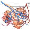
The adrenal glands are a pair body located above the pole of each kidney. They are referring to the glands of internal secretion. In the adrenal glands there is a cortical and brainstant. It is there that the hormones are produced adrenaline, norepinephrine and glucocorticosteroids. They take part in the work of the heart, regulating the tone of blood vessels and metabolism.
If the doctor during inspection will suspect adrenal insufficiency, then the next step will be the assignment of the ultrasound of adrenal glands. Indications for research:
- permanent thirst;
- pigment spots on the face;
- arterial hypertension;
- muscle weakness;
- involuntary muscle twitching;
- excess body weight;
- infertility;
- violation of the sexual system;
- cysts and adrenal tumors.

How to prepare for the procedure
To ensure the maximum reliable result of ultrasound research, it is necessary to follow certain rules for preparing for ultrasound. A few days before the procedure, you need to switch to a diet, providing for power, which will not provoke the formation of gases in the intestine and the fermentation process. A sharp, smoked, salted and fried food fall into the list of prohibited products. It is also necessary to failure from the use of carbonated and alcoholic beverages within 3 days before the procedure.
Vegetables, fruits, cereals, potatoes, low-fat meat and fish are allowed. From drinks is allowed black and green tea, coffee, compotes, juices. To prepare correctly, in the evening before the procedure, the enema should be conducted at home - so you can clean the intestines for a clearer ultrasonic picture. The study is made on an empty stomach, so on the day of the procedure it is necessary to refrain from meals, and on the eve of a light dinner allowed.
It is recommended to take with you mineral water Without gas, since in the study of adrenal glands, kidney and retroperitoneal space, a specialist will look a ureter and a bladder. The unfinished bladder is not visible by ultrasonic waves, so 500 ml fluid intake is mandatory.

Technique of ultrasound research
To carry out ultrasound research, the patient needs to lie down on the back or side and release the area of \u200b\u200bthe abdomen and the waist. The doctor then inflicts a special gel on the inspection site to ensure the best contact of the Uz-sensor with skin and eliminate the air between the skin and the device. The procedure takes up to 15 minutes.

Children allowed to carry out ultrasound diagnostics from birth. Newborns make research regardless of the fullness of the bladder. Before the procedure is allowed to receive a child medicinal preparationsreduced gas formation in the intestines.
Ultrasound adrenal glands
There are a number of features in the diagnosis of adrenal glands. The survey begin with the visualization of the right adrenal gland. To do this, the patient should go to the right side and take a deep breath. Assess the size of the adrenal gland, the presence of additional volume formations, the state of the cortical and brainstant, the presence of cyst or tumors.
Then go to the left. The procedure is similar, but the visualization process is hampered by the peculiarities of its location. You can not always see the adrenal gland on the ultrasound. Often, the procedure is combined with biochemical blood tests to clarify the full picture of the disease.
Ultrasound examination of the kidneys and retroperitoneal space
Two types of ultrasound of the kidneys are distinguished - echography and doppler. The first allows you to determine the form and sizes of the organ, the structure of the parenchyma, the presence of pathological inclusions. Dopplerography shows the changes and nature of blood flow in renal arteries. The kidneys are located at the level of the 12th breast - 1-2th lumbar vertebrae. The right below is left, since the liver presses on it. The length of the kidney is 10-12 cm. When breathing, these organs are able to shift by 2 cm.
An increase in the size of the kidneys according to the results of the ultrasound may indicate possible inflammation. Reducing, or wrinkling the body speaks of the presence of a dystrophic process. The presence of concrections in the kidney parenchyma or along the ureter, makes it possible to diagnose "Urolithiasis". Dopplerography speaks about the narrowing of renal arteries or veins thrombosis.
Ultrasonic research method helps the doctor to establish the correct diagnosis, assign adequate therapy and speed up the patient's recovery process.
Ultrasound of the abdominal organs and retroperitoneal space - the procedure is integrated, intended to diagnose diseases of several internal organs during one survey session. The ultrasound procedure is considered one of the safest research and informative medical methods. Allows you to evaluate the physical and functional state of the organs, to identify pathological changes at an early stage of their development.
What is the study directed?
Ultrasound of the retroper terrestrial space - a procedure aimed at studying the state of organs not covered with peritoneum or in its cavity and under its cover is only partially.
These include:
- kidney;
- adrenal glands;
- ureterals;
- lymph nodes;
- pancreas.
Most often, the Uz-diagnosis of the retroperitoneal space and the abdominal organs is carried out in conjunction in the framework of one procedure. Separately, a study of any organ for the appointment of the attending physician may be conducted.

 Ultrasound of the retroperitoneal space - a comprehensive study of organs not covered with the abdominal shell
 Where can I make an ultrasound of a barrier space organs?
- in specialized diagnostic clinics;
- in ambulatory;
- in hospitals;
- in private medical institutions.
Almost all medical institutions are equipped with ultrasound devices and keep in their staff professionals who can work with such equipment and study the state of the abdominal cavity and retroperitoneal space. This procedure has a high level of accessibility for the population, both in terms of its physical and financial accessibility.
Preparatory activities depend on the scope of research, its goals and a particular body, which is subject to study. It is not difficult to prepare for the ultrasound, no special measures are required.
Preparation is carried out in order to reduce the manifestations of meteorism. The accumulation of a large number of gases in the intestinal hinges makes it difficult to echolocation and affects the quality of the visualization of the organ.
To reduce gas formation, the intestines are recommended 2-3 days before the diagnosis to exclude from its dietary products causing gas production:
- milk;
- fresh vegetables and fruits;
- fresh bread and pastry baking;
- peas, beans, beans and lentils;
- fat and sharp dishes;
- carbonated drinks.
If the patient suffers from meteorism, you can take the winder and enterosorbent per day before the procedure. Before the procedure in the morning hours it is not recommended to eat. The last meal must take place no earlier than 8 hours before the ultrasound.
Preparation for the study of the bladder has a small feature. If you plan to study this organ, you should drink 500 ml of water 1 hour before the ultrasound and not to urinate before the procedure.
It is necessary to take a diaper, to store the couch, and napkins to remove excess gel. So all the preparations for ultrasound kidneys and adrenal glands, the hepatobiliary system and the retroperitoneal space.
How is an ultrasound study?
The procedure is carried out in the lying position (on the back, side, abdomen), sometimes to improve the visual picture - standing. When diagnosing the state of the kidneys, there will have to do a study in several positions. The patient should bargain the test zone. On the skin in the field of study, the diagnostician does not cause gel and through the sensor examines the organs and systems designated by the doctor. The duration of the procedure is up to 30 minutes. The procedure is not invasive, not traumatic, the patient does not deliver anxiety.
 During the ultrasound, the surveyed must be changed several times the position of the body for better visualization of the organ
 Contraindications for ultrasound diagnostics
Absolute contraindications ultrasound does not have, regardless of which organ should be explored (kidney, adrenal glands, pancreas and others). It is appointed pregnant women and breastfeeding. To date side Effects In the near or remote future, it was not revealed. Relative contraindications are inflammatory or purulent skin lesions or burns in the study area.
Testimony for ultrasound
- with the presence of appropriate complaints in the patient;
- under medical examination, including in preventive purposes;
- before surgery;
- for monitoring the results of surgical intervention;
- during pregnancy in the presence of testimony.
The pathology of the kidneys or adrenal glands can be suspected in the event of a persistent increase in blood pressure, accompanied by strong headaches and edema age. For persistent or frequent edema of the face and lower extremities, the cause is likely to be kidney disease. Pain in the lumbar region and violation of urine fat (change in the frequency of urination or volume of the dedicated liquid), a change in urine color is also characteristic of kidney problems.
The ultrasound diagnosis of the retroperitoneal space and organs located in the abdominal cavity is recommended:
- with complaints of the patient on pain in the top of the abdomen, the right hypochondrium or hazing pain;
- with a sense of gravity, meteorism, the feeling of cutting after meals.
This procedure allows you to diagnose serious diseases in the early stage process. These diseases include:
- pathology of kidney (urolithiasis, nephropathy, inflammatory diseases);
- diseases of adrenal glands (neoplasms, hyperplasia and hypoplasia).
Ultrasound informative for diseases of vascular genesis (aneurysms, hemangiomas). And with the pathology of the lymph nodes of an inflammatory nature or in the expanding of metastasis.
Diagnostic
Usually, in conclusion, an ultrasound procedure, a doctor indicates not only an organ pathology and its degree, but also lesions (if any) of the abdominal wall tissues and other pathologies found in the retroperitoneal space. It may be tumors, infiltrates or abscesses.
When describing the abdominal cavity organs and the retroperitoneal space, the doctor assesses their location, clarity of the contours, form, structure, volume. If foreign inclusions, tumors, ultrasonic cysts indicate their location, sizes, connection with the abdominal wall, vascular plexus or lymph nodes.
How do kidney ultrasound do?
- infertility in women;
Preparing for research:
Ultrasound of the kidneys - a simple, accessible study based on the effects of ultrasound on the soft tissue of a person. Since the method is not associated with X-ray radiation, it is not only effective and accurate, but also absolutely not dangerous and painless for the patient.
Indications for the ultrasound of the kidneys:
- Pain in the lower back and suspected tumor;
- Suspicion of kidney inflammation (glomerulonephritis, pyelonephritis);
- Suspicion of urolithiasis;
- Pathology of the development of kidneys or vessels;
- With increasing body temperature for a long time and without an explicit reason.
With the help of ultrasound research, such diseases can be identified as:
- Chronic pyelonephritis is inflammation of the kidney tissue, in which the outflow of urine is disturbed. Pregnant women are particularly susceptible to this disease. That is why, during pregnancy, in the event of pains in the lower back, an ultrasound is appointed.
- Urolithiasis - is the process of formation of kidney stones;
- Anomalies of renal vessels are defects of renal veins and arteries. Manifest themselves in the form of additional veins and arteries, as well as the narrowing of the renal artery, which deprives the kidney of a sufficient number of blood coming into it;
- Cysta - cavity with liquid. It can be both single and multiple (polycystic disease - a congenital disease characterized by the presence of a cyst in both kidneys);
- Tumors - both malignant and benign neoplasms;
- Piilyectasia - the expansion of the buds of the kidneys (there is congenital and acquired). Often does not require treatment and passes with age.
Contraindications for conducting research - no.
- the appearance of pigmentation on the skin;
- feeling a patient constant weakness;
- obesity, while unknown the reason for his appearance;
- infertility in women;
- the appearance of multiple stretch marks on the skin;
- reduction or increase in pressure on obscure reasons;
- deviations that indicate the presence of body tumors.
- Hyperplasia - appears even before birth, when the baby is in the abdomen of Mom.
- Inflammatory changes and hematomy arise when the abdomen is closed; Cysts. There are rarely found.
- Tumors. May be benign (adenoma), but also malignant (sarcoma). In most cases, adenomas are found. They exist asymptomatic and do not cause harm to the body. Sarcoma is rarely detected.
- Pain in the lower back and suspected tumor;
- Suspicion of kidney inflammation (glomerulonephritis, pyelonephritis);
- Suspicion of urolithiasis;
- Pathology of the development of kidneys or vessels;
- With increasing body temperature for a long time and without an explicit reason.
- Chronic pyelonephritis is inflammation of the kidney tissue, in which the outflow of urine is disturbed. Pregnant women are particularly susceptible to this disease. That is why, during pregnancy, in the event of pains in the lower back, an ultrasound is appointed.
- Urolithiasis - is the process of formation of kidney stones;
- Anomalies of renal vessels are defects of renal veins and arteries. Manifest themselves in the form of additional veins and arteries, as well as the narrowing of the renal artery, which deprives the kidney of a sufficient number of blood coming into it;
- Cysta - cavity with liquid. It can be both single and multiple (polycystic disease - a congenital disease characterized by the presence of a cyst in both kidneys);
- Tumors - both malignant and benign neoplasms;
- Piilyectasia - the expansion of the buds of the kidneys (there is congenital and acquired). Often does not require treatment and passes with age.
Preparation for the ultrasound of the kidneys:
The survey is not conducted on an empty stomach, so it is not necessary to be offered from meals and drink. The only difficulty in conducting the procedure may be increased gas formation in the intestine. Therefore, the preparation of the ultrasound of the kidneys will be that 3 days before the survey should be abandoned from solid milk, raw vegetables and fruit, black bread. In addition, it is also recommended to take "Espumizan" or activated carbon. If the patient has a violation of digestion, the doctor may appoint additionally "mesim" or "festal".
How do kidney ultrasound do?
Ultrasound kidneys is carried out in a state lying. The patient is located on the couch, face to the doctor - diagnostic (lying on the back). As with ultrasound of any other internal organA special colorless gel is applied to the area under study, which improves the passage of ultrasound through soft tissues. The doctor then using an ultrasound sensor produces scanning, and the resulting result in the form of a black and white photo is displayed on the computer monitor screen. The procedure lasts 15-20 minutes, the result is ready almost immediately.
Adrenal glands - paired organs located above the kidneys. It performs the role of the glands of the internal secretion. Functions are in regulating metabolism, the production of hormones needed to ensure important life processes
Indications for Uzi adrenal glands:
Ultrasound of adrenal glands can identify the following organs of organs:
Contraindications for research - no.
Preparing for research:
All the preparation of the ultrasound of the adrenal glands is not complicated and includes, most likely, recommendations than requirements. A couple of days before the procedure to comply with a slicing diet, eliminating products leading to increased formation of gases (coarse vegetables, smoked salinity, salinity, sausages), as well as fried and oily food and meat. It is allowed to eat vegetables (boiled, stewed), fruits, drink non-carbonated juices. In the evening before the procedure, drink a laxative. On an empty stomach procedure.
Before ultrasonic diagnostics, pass the blood test to hormones and visit the endocrinologist.
Ultrasound of the kidneys - a simple, accessible study based on the effects of ultrasound on the soft tissue of a person. Since the method is not associated with X-ray radiation, it is not only effective and accurate, but also absolutely not dangerous and painless for the patient.
Indications for the ultrasound of the kidneys:
With the help of ultrasound research, such diseases can be identified as:
Contraindications for conducting research - no.
The abdominal cavity of a person from the inside wore a thin shell called the peritoneum, providing the selection and absorption of a minor amount of fluid for better work all organs. However, there are organs that this shell does not affect: they are for the peritoneum. That is why the space limited in front of the peritinous, and behind the lumbar muscles and the spine, is called retroperitoneal, or retroperitoneal. The study of it with ultrasound is often included in the standard protocol and is carried out along with the ultrasound of the abdominal organs.
A little anatomy
To understand where the retroperitoneum is localized, you just need to know where the lumbar back area is. Now you can accurately name the organs located in the retroperitoneal space:
- kidneys with ureters;
- adrenal glands;
- aorta and lower hollow vein passing along the spine.
There are organs that are partially covered with peritoneum and are in the abdominal cavity, and the other department is located retroinchenously. These authorities include:
- pancreas;
- duodenum;
- part of the large intestine: ascending and downward colon.
In addition to organs, the retroperitoneal space is filled with fatty tissue that performs the supporting function.
Ultrasound procedure
Ultrasound of the retroperitoneal space today is one of the most accessible methods for the diagnosis of kidney pathology, adrenal glands. The inspection of the vessels, the pancreas and the intestines is included in the study of the abdominal organs, however, according to emergency testimony, you can make a sonography of any structure whose pathology suspects a doctor, up to the soft tissues of the lumbar region in suspected the presence of hematoma. Retroperitoneum inspect the following testimony:

Preparation for ultrasound
Depending on which an organ or system, it is necessary to make an emphasis, preparation for the procedure is somewhat different.
Common is that it is necessary to take a pellery on which it will be possible to lie during the procedure and wipe the remnants of the gel after it. Some medical organizations provide disposable pelleys, but it is worth getting their towel to suffer. It is necessary to take into account that wet wipes are not very good to apply in this case, as they are poorly collecting gel that remains on the skin.
Urinary system
No special preparatory activities are required. However, you should pay attention to drinking mode: you should not drink a lot before ultrasound, as it will provoke the active working of the kidneys and can lead to incorrect interpretation of some indicators when inspection. For example, Lohanka Kidneys will expand slightly, according to which the urine from the kidneys is going to the ureter and further into the bladder.
Expanded Lohanka Kidney can talk about the presence of pathology or about the normal physiological process.
Adrenal glands
They are a pair endocrine organ located near the upper poles of the kidneys. The tissue of adrenal glands with ultrasound is practically not visible, so the doctor visually evaluates the zone of their location, in which any clearly defined additional education, if they are.

The zone of the right adrenal gland is visible better, and the zone of the left visualize is more difficult. This is due to the peculiarity of the anatomical location of the adrenal and neighboring organs themselves. A stomach arrives to the left adrenal gland, so the study is conducted on an empty stomach.
On an empty stomach - this means that it is impossible to eat and drink 8 hours before the study, because both solid, and liquid food will interfere with the inspection.
Aorta and lower hollow vein
For the examination of vessels, a diet is needed with the exception of products that promote fermentation and gas formation in the intestine, as well as the reception of such medicines as:
- activated carbon or other enterosorbenta;
- enzyme preparations, for example, mezim, festal, pancreatin and others;
- waterproof agents: Siemeticone and its counterparts.
Conducting an ultrasound study of retroperitoneal space
Before the start of the inspection, it is necessary to free the research area from clothing, go to the couch, pre-shined the pelleny, and perform the instructions of a specialist who will apply a gel on the research area or directly to the sensor and start examining.
You need to be prepared for the fact that during the inspection you will have to change the position of the body several times. If the aorta can be explored in the lying position on the back, then the kidneys and adrenal glands need to be examined on all sides, that is, in the position of lying on the back, on the side, on the stomach, sitting and standing.
Normal indicators and most frequent pathology
A qualitative study of the retroperitoneal space with ultrasound is impossible without defining the norm.
Kidney
The form of normal kidney oval or beanoid, contour is clear and smooth, sometimes wavy. The longitudinal size should not exceed 12 cm and be less than 10 cm. However, the size of the kidneys depends on the constitutional characteristics of a person and from the kind of activity, for example, professional athletes kidney may be greater.
The echostructure must be homogeneous, echogenicity of moderate or normal, that is, the kidney parenchyma is a little darker liver when ultrasound. The center of the kidney on the contrary, looks white.
Diffuse changes in kidneys
There is a change in the echostructure and echogenicity of the parenchyma of one or both kidneys.

Focal Pathology
The most frequent education detectable in ultrasound kidneys is cysts. They may be single and multiple, minor and giant, round and irregular shape. Small cysts need to be observed, that is, to inspect once a year. Very large sizes are cleaned.
Urolithiasis disease
Pathology of the kidneys characterized by the formation of various compositions in cups or lochank. When studying stones look like a bright white structure, which gives a black shadow. They can be multiple or isolated, small or large, round, oval or irregular shape.
Adrenal glands
Normally, this pair body is not visualized.
When conducting an ultrasound of the retroperitoneal space, focal changes in the adrenal glands are most often identified, about the nature of which is quite difficult to judge, so the method of choice is a computer or magnetic resonance imaging.
Aorta
The normal aortic diameter is about 25 mm if the expansion of the vessel section of the vessel with a dimmeter of more than 30 mm was revealed during the inspection, therefore, they talk about aneurysm.
The doctor also draws attention to the walls of the aorta, since atherosclerotic plaques are often detected in age patients.
If there is a need for an ultrasound of the progenitarian space, it is not necessary to pull, since there are vital organs in the retroperitoneum: kidneys, adrenal glands and the two largest organism vessels.