The cell wall is a rigid and dense membrane located above the cytoplasmic membrane. This element is characteristic of the cells of bacteria, fungi and plants. In addition to protecting the cell, the rigid shell also performs a number of other equally important functions.
Cell wall: general information
The cell wall of every organism has a number of features. For example, in bacteria, it consists mainly of murein. By the way, bacterial strains are divided into two types - gram-positive and gram-negative - precisely because of the structural features of the hard shell. This determines their sensitivity to antibiotics.
If we talk about the cell walls of fungi, then their main components are chitin and glucans. But the shells of algae can consist of different polysaccharides - mainly glucose and its compounds. By the way, the composition of the cell wall of algae is a very important taxon. It is worth remembering about the group whose representatives synthesize their own silica wall.
Plant cell wall and its functions
The principles of the structure of the rigid cell membrane are most conveniently studied by example And although mechanical protection is one of the most important, it is of much greater importance:
- provides mechanical and chemical resistance of the cell;
- prevents cell rupture in a hypotonic environment;
- the cell wall is also an ion exchanger, since ions are absorbed and released through it;
- takes part in the transport of organic compounds.
Cell wall structure
It is customary to distinguish three main components in the plant wall: the frame, the matrix and the encrusting substances.
The cell wall of a plant is composed of cellulose. Due to the formation of cellulose molecules, they form strong microfibrils, which are immersed in the base substance, or matrix.
The matrix of the cell wall makes up approximately 60% of its total mass. It fills the space between microfibrils, and also creates strong bonds between macromolecules, provides elasticity and strength of this cellular structure. The main components of the matrix are hemicellulose and pectin.
- Hemicellulose is a polysaccharide similar in structure to cellulose, but with shorter and more branched monomer chains.
- also belong to polysaccharides, but they also contain residues. Due to the formation of chemical bonds with calcium and magnesium ions, pectin takes part in the formation of the middle plates - the places where two neighboring cells connect to each other. By the way, a large amount of pectin is found in plant fruits.
The encrusting substances in most cases are represented by lignin, which makes up about 30% of the dry mass of the cell wall.
- Lignin can be deposited both as a continuous layer and in the form of individual elements - spirals, nets, or rings. This substance acts like cement - it holds the cellulose fibers together. Due to lignification, the cell wall becomes more resistant and less permeable to water. By the way, it is lignin that is responsible for lignification of plants.
Quite often, substances such as cutin, suberin and wax are deposited on the outer surface of the cell membrane.
Suberin is deposited on the inner side of the cell membrane, providing a corking process. Such a cell becomes absolutely impervious to moisture, so its contents quickly die off, and the free space is filled with air.
The main function of wax substances and cuticles is to protect cells from infection, as well as to reduce the level of water evaporation.
We can say that the cell wall is a very important element of the plant cell, which ensures its normal development.
At the dawn of the development of life on Earth, all cellular forms were represented by bacteria. They sucked in organic matter dissolved in the primary ocean through the surface of the body.
Over time, some bacteria have adapted to produce organic matter from inorganic matter. To do this, they used the energy of sunlight. The first ecological system arose in which these organisms were producers. As a result, oxygen appeared in the Earth's atmosphere, released by these organisms. With its help, much more energy can be obtained from the same food, and the additional energy can be used to complicate the structure of the body: dividing the body into parts.
One of the important achievements of life is the separation of the nucleus and cytoplasm. The core contains hereditary information. A special membrane around the core made it possible to protect against accidental damage. As necessary, the cytoplasm receives commands from the nucleus that direct the vital activity and development of the cell.
Organisms in which the nucleus is separated from the cytoplasm have formed a super-kingdom of nuclear (these include plants, fungi, animals).
Thus, the cell - the basis of the organization of plants and animals - arose and developed in the course of biological evolution.
Even with an unaided eye, and even better under a magnifying glass, you can see that the pulp of a ripe watermelon consists of very small grains, or grains. These are cells - the smallest “bricks” that make up the bodies of all living organisms, including plant ones.
The life of a plant is carried out by the combined activity of its cells, which create a single whole. With the multicellularity of plant parts, there is a physiological differentiation of their functions, the specialization of various cells depending on their location in the plant body.
A plant cell differs from an animal cell in that it has a dense shell that covers the inner contents from all sides. The cell is not flat (as it is usually portrayed), it most likely looks like a very small vesicle filled with mucous contents.
Plant cell structure and function
Let's consider a cell as a structural and functional unit of an organism. Outside, the cell is covered with a dense cell wall, in which there are thinner areas - pores. Under it is a very thin film - a membrane that covers the contents of the cell - the cytoplasm. There are cavities in the cytoplasm - vacuoles filled with cell sap. In the center of the cell or near the cell wall there is a dense body - a nucleus with a nucleolus. The nucleus is separated from the cytoplasm by a nuclear envelope. Small bodies - plastids - are distributed throughout the cytoplasm.
Plant cell structure
The structure and function of plant cell organelles
| Organoid | Drawing | Description | Function | Peculiarities |
Cell wall or plasma membrane | Colorless, transparent and very durable | It allows substances into and out of the cell. | The cell membrane is semi-permeable |
|
Cytoplasm | Thick viscous substance | All other parts of the cell are located in it. | Is in constant motion |
|
The nucleus (an important part of the cell) | Rounded or oval | Provides the transfer of hereditary properties to daughter cells during division | Central part of the cell |
|
Spherical or irregular shape | Takes part in protein synthesis | |||
 | A reservoir separated from the cytoplasm by a membrane. Contains cell juice | Reserve nutrients and waste products are accumulated that are unnecessary for the cell. | As the cell grows, small vacuoles merge into one large (central) vacuole |
|
Plastids | Chloroplasts | Harness the light energy of the sun and create organic from inorganic | The shape of the discs delimited from the cytoplasm by a double membrane |
|
Chromoplasts | Formed as a result of the accumulation of carotenoids | Yellow, orange or brown |
||
 | Leukoplasts | Colorless plastids | ||
Nuclear shell | Consists of two membranes (outer and inner) with pores | Separates the nucleus from the cytoplasm | Enables exchange between the nucleus and the cytoplasm |
The living part of the cell is a membrane-limited, ordered, structured system of biopolymers and internal membrane structures that participate in a set of metabolic and energy processes that maintain and reproduce the entire system as a whole.
An important feature is that there are no open membranes with free ends in the cell. Cell membranes always enclose cavities or areas, covering them from all sides.

Modern generalized plant cell diagram
Plasmalemma(outer cell membrane) - ultramicroscopic film 7.5 nm thick, consisting of proteins, phospholipids and water. It is a very elastic film that is well wetted with water and quickly restores its integrity after damage. It has a universal structure, i.e., typical for all biological membranes. In plant cells, outside the cell membrane, there is a strong cell wall that provides external support and maintains the shape of the cell. It consists of fiber (cellulose), a water-insoluble polysaccharide.
Plasmodesmata plant cells, are submicroscopic tubules that penetrate the membranes and are lined with a plasma membrane, which thus passes from one cell to another without interruption. With their help, the intercellular circulation of solutions containing organic nutrients occurs. They are also used to transfer biopotentials and other information.
Pore called holes in the secondary membrane, where the cells are separated only by the primary membrane and the median plate. The areas of the primary membrane and the median lamina separating adjacent pores of adjacent cells are called the pore membrane or pore closing film. The closure film of the pores penetrates the plasmodesmenable tubules, but a through hole is usually not formed in the pores. The pores facilitate the transport of water and solutes from cell to cell. In the walls of neighboring cells, as a rule, one against the other, pores are formed.
Cell membrane has a well-defined, relatively thick shell of a polysaccharide nature. The plant cell membrane is a product of the activity of the cytoplasm. The Golgi apparatus and the endoplasmic reticulum are actively involved in its formation.

Cell membrane structure
The basis of the cytoplasm is its matrix, or hyaloplasm, a complex colorless, optically transparent colloidal system capable of reversible transitions from sol to gel. The most important role of hyaloplasm is to unite all cellular structures into a single system and to ensure interaction between them in the processes of cellular metabolism.
Hyaloplasm(or matrix of the cytoplasm) constitutes the internal environment of the cell. Consists of water and various biopolymers (proteins, nucleic acids, polysaccharides, lipids), of which the bulk are proteins of various chemical and functional specificity. The hyaloplasm also contains amino acids, monosaccharides, nucleotides and other low molecular weight substances.
Biopolymers form a colloidal medium with water, which, depending on the conditions, can be dense (in the form of a gel) or more liquid (in the form of a sol), both in the entire cytoplasm and in its individual areas. In the hyaloplasm, various organelles and inclusions are localized and interact with each other and the environment of the hyaloplasm. Moreover, their location is most often specific to certain types of cells. Through the bilipid membrane, the hyaloplasm interacts with the extracellular environment. Consequently, hyaloplasm is a dynamic environment and plays an important role in the functioning of individual organelles and the vital activity of cells in general.
Cytoplasmic formations - organelles
Organelles (organelles) are structural components of the cytoplasm. They have a certain shape and size, and are mandatory cytoplasmic structures of the cell. In their absence or damage, the cell usually loses the ability to continue to exist. Many of the organelles are capable of division and self-reproduction. Their dimensions are so small that they can only be seen through an electron microscope.
Core
The nucleus is the most visible and usually the largest organelle of the cell. It was first explored in detail by Robert Brown in 1831. The nucleus provides the most important metabolic and genetic functions of the cell. It is quite variable in shape: it can be spherical, oval, lobed, lenticular.
The nucleus plays a significant role in the life of the cell. The cell from which the nucleus was removed no longer secretes a membrane, it ceases to grow and synthesize substances. The products of decay and destruction increase in it, as a result of which it quickly dies. The formation of a new nucleus from the cytoplasm does not occur. New nuclei are formed only by fission or crushing of the old one.
The inner content of the nucleus is the karyolymph (nuclear juice), which fills the space between the structures of the nucleus. It contains one or more nucleoli, as well as a significant number of DNA molecules connected to specific proteins - histones.

Nucleus structure
Nucleolus
The nucleolus, like the cytoplasm, contains mainly RNA and specific proteins. Its most important function is that it forms ribosomes, which carry out the synthesis of proteins in the cell.
Golgi apparatus
The Golgi apparatus is an organoid that is universally distributed in all types of eukaryotic cells. It is a multi-tiered system of flat membrane sacs, which thicken along the periphery and form vesicular processes. It is most often located near the nucleus.

Golgi apparatus
The Golgi apparatus necessarily includes a system of small vesicles (vesicles), which are detached from thickened cisterns (discs) and are located along the periphery of this structure. These vesicles play the role of an intracellular transport system of specific sector granules and can serve as a source of cellular lysosomes.
The functions of the Golgi apparatus are also in the accumulation, separation and excretion outside the cell with the help of bubbles of the products of intracellular synthesis, decay products, and toxic substances. The products of the synthetic activity of the cell, as well as various substances entering the cell from the environment through the channels of the endoplasmic reticulum, are transported to the Golgi apparatus, accumulate in this organoid, and then in the form of droplets or grains enter the cytoplasm and are either used by the cell itself, or are excreted outside ... In plant cells, the Golgi apparatus contains enzymes for the synthesis of polysaccharides and the polysaccharide material itself, which is used to build the cell wall. It is believed to be involved in the formation of vacuoles. The Golgi apparatus was named after the Italian scientist Camillo Golgi, who first discovered it in 1897.
Lysosomes
Lysosomes are small vesicles bounded by a membrane, the main function of which is to carry out intracellular digestion. The use of the lysosomal apparatus occurs during the germination of the plant seed (hydrolysis of reserve nutrients).
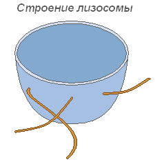
Lysosome structure
Microtubules
Microtubules are membrane, supramolecular structures consisting of protein globules arranged in spiral or rectilinear rows. Microtubules perform primarily a mechanical (motor) function, providing mobility and contractility of cell organelles. Located in the cytoplasm, they give the cell a certain shape and ensure the stability of the spatial arrangement of organelles. Microtubules facilitate the movement of organelles to places that are determined by the physiological needs of the cell. A significant number of these structures are located in the plasmalemma, near the cell membrane, where they are involved in the formation and orientation of cellulose microfibrils of plant cell membranes.

Microtubule structure
Vacuole
Vacuole is the most important component of plant cells. It is a kind of cavity (reservoir) in the mass of the cytoplasm, filled with aqueous solution mineral salts, amino acids, organic acids, pigments, carbohydrates and separated from the cytoplasm by the vacuolar membrane - tonoplast.
The cytoplasm fills the entire internal cavity only in the youngest plant cells. With the growth of the cell, the spatial arrangement of the initially continuous mass of cytoplasm changes significantly: small vacuoles filled with cell juice appear in it, and the entire mass becomes spongy. With further growth of the cell, individual vacuoles merge, pushing back to the periphery the layer of cytoplasm, as a result of which there is usually one large vacuole in the formed cell, and the cytoplasm with all organelles are located near the membrane.
Water-soluble organic and mineral compounds of vacuoles determine the corresponding osmotic properties of living cells. This solution of a certain concentration is a kind of osmotic pump for regulated penetration into the cell and the release of water, ions and metabolite molecules from it.
In combination with a layer of cytoplasm and its membranes, which are characterized by semipermeability properties, the vacuole forms an effective osmotic system. Such indicators of living plant cells as osmotic potential, sucking force and turgor pressure are osmotically determined.

Vacuole structure
Plastids
Plastids are the largest (after the nucleus) cytoplasmic organelles, inherent only in the cells of plant organisms. They are not found only in mushrooms. Plastids play an important role in metabolism. They are separated from the cytoplasm by a double membrane membrane, and some of their types have a well-developed and ordered system of internal membranes. All plastids are of the same origin.
Chloroplasts- the most widespread and most functionally important plastids of photoautotrophic organisms, which carry out photosynthetic processes that ultimately lead to the formation of organic matter and the release of free oxygen. Chloroplasts of higher plants have a complex internal structure.

Chloroplast structure
The sizes of chloroplasts in different plants are not the same, but their average diameter is 4-6 microns. Chloroplasts are able to move under the influence of the movement of the cytoplasm. In addition, under the influence of illumination, an active movement of amoeba-type chloroplasts towards the light source is observed.
Chlorophyll is the main substance of chloroplasts. Thanks to chlorophyll, green plants are able to use light energy.
Leukoplasts(colorless plastids) are clearly marked cytoplasmic bodies. Their size is somewhat smaller than the size of chloroplasts. More and more monotonous and their shape, closer to spherical.
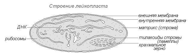
Leukoplast structure
Found in the cells of the epidermis, tubers, rhizomes. When illuminated, they very quickly turn into chloroplasts with a corresponding change in the internal structure. Leukoplasts contain enzymes, with the help of which starch is synthesized from excess glucose formed in the process of photosynthesis, the bulk of which is deposited in storage tissues or organs (tubers, rhizomes, seeds) in the form of starch grains. In some plants, fats are deposited in leukoplasts. The reserve function of leukoplasts occasionally manifests itself in the formation of storage proteins in the form of crystals or amorphous inclusions.
Chromoplasts in most cases, they are derivatives of chloroplasts, occasionally leukoplasts.

Chromoplast structure
Ripening of rose hips, peppers, tomatoes is accompanied by the transformation of chloro- or leukoplasts of pulp cells into carotenoidoplasts. The latter contain mainly yellow plastid pigments - carotenoids, which, when ripe, are intensively synthesized in them, forming colored lipid droplets, solid globules or crystals. In this case, chlorophyll is destroyed.
Mitochondria
Mitochondria are organelles that are characteristic of most plant cells. They have a changeable shape of sticks, grains, threads. Discovered in 1894 by R. Altman using a light microscope, and the internal structure was studied later using an electron microscope.
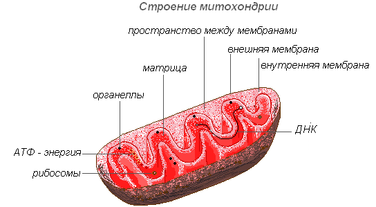
Mitochondrion structure
Mitochondria have a two-membrane structure. The outer membrane is smooth, the inner one forms outgrowths of various shapes - tubules in plant cells. The space inside the mitochondrion is filled with a semi-liquid content (matrix), which includes enzymes, proteins, lipids, calcium and magnesium salts, vitamins, as well as RNA, DNA and ribosomes. The enzymatic complex of mitochondria accelerates the complex and interconnected mechanism of biochemical reactions that result in the formation of ATP. In these organelles, the cells are provided with energy - the transformation of the energy of chemical bonds of nutrients into high-energy ATP bonds in the process of cellular respiration. It is in the mitochondria that the enzymatic breakdown of carbohydrates occurs, fatty acids, amino acids with the release of energy and its subsequent transformation into energy ATP. The accumulated energy is spent on growth processes, on new syntheses, etc. Mitochondria multiply by fission and live for about 10 days, after which they undergo destruction.
Endoplasmic reticulum
The endoplasmic reticulum is a network of channels, tubules, vesicles, cisterns located inside the cytoplasm. Discovered in 1945 by the English scientist K. Porter, it is a system of membranes with an ultramicroscopic structure.
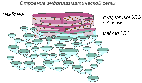
The structure of the endoplasmic reticulum
The entire network is integrated into a single whole with the outer cell membrane of the nuclear envelope. Distinguish between smooth and rough EPS, bearing ribosomes. On the membranes of smooth EPS there are enzyme systems involved in adipose and carbohydrate metabolism... This type of membrane predominates in seed cells rich in storage substances (proteins, carbohydrates, oils), ribosomes attach to the membrane of granular EPS, and during the synthesis of a protein molecule, the polypeptide chain with ribosomes is immersed in the EPS channel. The functions of the endoplasmic reticulum are very diverse: transport of substances both inside the cell and between neighboring cells; division of the cell into separate sections, in which various physiological processes and chemical reactions take place simultaneously.
Ribosomes
Ribosomes are non-membrane cellular organelles. Each ribosome consists of two particles that are not the same size and can be divided into two fragments, which continue to retain the ability to synthesize protein after combining into a whole ribosome.

Ribosome structure
Ribosomes are synthesized in the nucleus, then leave it, passing into the cytoplasm, where they attach to the outer surface of the membranes of the endoplasmic reticulum or are located freely. Depending on the type of the synthesized protein, ribosomes can function alone or combine into complexes - polyribosomes.
The cell wall is a protoplast derivative, i.e. formed in the course of its life (Fig. 61). It gives the cell a definite shape, protects the protoplast and, by resisting intracellular pressure, prevents cell rupture. Carrying out the functions of the internal skeleton of a plant, the cell walls give its organs the necessary mechanical strength.
Cell walls transmit sunlight well, water and minerals dissolved in it easily move along them. Between the walls of neighboring cells there is median plate - the pectin layer, which, in fact, is an intercellular substance, holds the walls of neighboring cells together. In those places where the cell walls of neighboring cells do not close, filled with water are formed intercellular spaces. The process of destruction of the intercellular substance, as a result of which the walls of neighboring cells are separated, is called maceration. Natural maceration can be observed
Rice. 61.
A- a diagram of the structure of the cell wall; B- scheme of the participation of the Golgi apparatus in the construction of the cell wall; V- detailed structure of the cell wall: 1 - median plate; 2 - it's time; 3 - secondary wall;
- 4 - primary wall; 5 - dictyosome; 6 - Golgi bubbles;
- 7- plasmalemma; 8- cell wall; 9- macrofibril;
- 10- microfibril; 11 - micelle; 12 - cellulose molecule;
- 13 - structure of a fragment of a cellulose molecule
in overripe fruits of apple, mountain ash, melon, etc. Artificial maceration is carried out, for example, when soaking flax stalks to separate bast fibers from them; it also occurs during heat treatment of fruits.
The cell wall contains polysaccharides: pectins, hemi-cellulose and cellulose. Very long cellulose molecules are arranged in an orderly parallel to each other (40-60 each), forming micelles. Micelles are collected in bundles - microfibrils, representing the main structural unit of cellulose. Microfibrils, in turn, are combined into macrofibrils- very thin fibers of indeterminate length. Cellulose macrofibrils are immersed in a highly watered matrix, consisting of pectins, hemicelluloses and some other substances. The strength of the cell wall is given by elastic microfibrils of cellulose, which are close in tensile strength to steel. The strength and elasticity of the cell wall underlies its ability to stretch reversibly. Thanks to pectins and hemicellulose, the cell wall is highly permeable to water - water and substances dissolved in it easily move along it from cell to cell.
The cell wall is adjacent to the outside of the plasmalemma, which is actively involved in its growth. Molecules of pectins, hemicellulose, cellulose and other substances are synthesized and accumulated in the cisterns of the dictyosomes of the Golgi apparatus. Golgi vesicles deliver them to the periphery of the protoplast - to the plasmalemma. At the point of contact between the bubble and the plasmalemma, the latter dissolves, and the contents of the bubble, being outside the plasmalemma, go to the construction of the cell wall. The bubble membrane not only restores the integrity of the plasma membrane, but also ensures its surface growth. The growth of the cell wall is carried out due to the enzymatic activity of the plasmalemma.
The walls of dividing and growing cells are called primary. They contain a lot of water (60-90%), pectins and hemicellulose predominate in their dry matter - no more than 30% cellulose. During cell division in the telophase of mitosis, the maternal cell is divided into two daughter cells as a result of the formation of a septum in its equatorial plane - the median plate. On both sides of the median plate, each of the two daughter cells begins to create its own primary cell wall. The growth of the median plate and primary walls of two daughter cells proceeds in a centrifugal direction - from the center of the mother cell to its periphery. The median lamina is very thin and consists of pectin.
The new cell formed as a result of division begins to grow, while its volume can increase by a factor of 100 or more. Cell growth proceeds mainly through stretching by absorbing water and increasing the volume of vacuoles. The resulting internal pressure stretches the primary wall, into which micelles of cellulose, pectin, and hemicellulose are easily incorporated. The way the cell wall grows by introducing building material between the existing structures is called Intussusception.
In the primary cell wall, there are initially thinner areas where cellulose fibrils are located more loosely, - primary pore fields. The primary pore fields of the walls of two adjacent cells usually coincide. Here, from one cell to another, the tubules of the endoplasmic reticulum pass - plasmodesmata. The paths by which plasmodesmata pass from one cell to another are called plasmodesmal tubules. Through these tubules, the hyaloplasms of neighboring cells are also connected. Intercellular transport of substances (hormones, amino acids, ATP, sugars, etc.) is carried out along the plasmodesmata. The protoplasts of the body cells, united into a single whole with the help of plasmodesm, are called symplasts. The transport of substances through the plasmodesmata is called symplastic.(The collection of cell walls, median lamina and intercellular spaces is called apoplast, walking along them apoplastic transport of substances.)
After the completion of cell growth, its wall can remain thin primary (in cells of educational tissues) or begin to grow in thickness (in cells of permanent tissues). The growth of the cell wall in thickness is called secondary thickening. As a result, it is deposited on the inner surface of the primary wall secondary wall that grows by appositions- the imposition of cellulose micelles on the already existing wall. In this case, the youngest layers of the secondary cell wall are located next to the plasmalemma. The secondary cell wall mainly performs supporting, mechanical functions. Its composition is much less water than in the primary, and cellulose predominates in dry matter (up to 50%). For example, in the secondary walls of unicellular hairs of cotton and bast fibers of flax, the cellulose content can reach 95%.
Secondary thickening of the cell wall occurs unevenly. Areas of the secondary cell wall at the locations of the primary pore fields usually remain non-thickened. Such non-thickened areas of the cell wall are called pores. The pores in the walls of two adjacent cells, as a rule, coincide, forming a couple of pores. The pore channel formed by a pair of pores is blocked pore closing film - a septum consisting of a median plate and two primary walls of adjacent cells. The closing film of the pore is permeated by numerous plasmodesmal tubules through which plasmodesmata pass.
Distinguish between pores simple and edged(fig. 62). In simple pores, the diameter of their section of the pore channel is the same along the entire length, i.e. it is cylindrical in shape. Simple pores are typical of parenchymal cells. Bordered pores are characteristic of the walls of cells that conduct water with dissolved minerals - tracheids and vascular segments. At such pores, their portion of the pore channel has the shape of a funnel, which with its wide side adjoins the closing film of the pore.
In the cells of conductive tissues of conifers, the closing film of the pore is permeable to water only at the edges, since its central disc-shaped thickened and lignified part is torus - does not allow water to pass through. Torus acts as a valve. If the water pressure in neighboring cells is not the same, the closing film is deflected and the torus blocks the movement of water along the pore channel.

Rice. 62.
A- simple; B- bordered; V- semi-bordered:
1 - closing film; 2 - pore channel; 3 - torus
In the walls of water-conducting cells, in addition to pores, perforations- through holes (vascular segments, water-storing cells of sphagnum moss).
Cell wall modifications. Depending on the functions performed by the cell, its wall can be modified due to the deposition of any substances in it. Its usual modifications are lignification, suberization, cutinization, mineralization and mucousness.
Lignification of the cell wall, or lignification, occurs as a result of the deposition of lignin in the intermicellar spaces - a substance of an aromatic nature with a complex chemical structure. At the same time, the strength and hardness of the wall increase, but its elasticity decreases. Lignified walls are able to pass water and air. With a lignified cell wall, the protoplast of the cell can remain alive, but usually dies off. Some woody plants accumulate up to 30% of lignin in the wood. Lignin can also accumulate in the cell walls of aging grass shoots, which significantly reduces their feed value and determines the timing of hay harvesting. In the process of obtaining cellulose pulp from wood, which is necessary for the production of paper, artificial lignification is carried out. Natural lignification of the cell wall is possible, but rare.
Corking, or suberinization, - deposition in the cell wall of a persistent fat-like amorphous substance suberin (hydrophobic polymer). The corked cell walls are impermeable to gases and water, which causes the death of the protoplast. Cork-walled cells reliably protect plants from water loss, extreme temperatures, pathogenic bacteria and fungi.
Cutinization - deposition in the cell walls of cutin (a substance similar in chemical composition to Suberin). Cutin is usually deposited in the surface layers of the outer walls of cells and on their surface. In the form of a film - cuticle - it covers, for example, the surface of the cells of the integumentary tissue - the epidermis.
Mineralization the cell wall occurs due to the deposition of calcium and silica salts in it. These substances impart hardness and brittleness to the wall. The process of mineralization in the cell walls of the epidermis of shoots of cereals, sedges, and horsetails is especially pronounced. For this reason, it is recommended to mow the shoots of sedges and grasses before they bloom - later, due to strong mineralization, they coarse, which reduces the quality of the hay.
Slime- the transformation of cellulose and cell wall pectins into special polysaccharides - mucus and gums, capable of strong swelling when in contact with water. Slicking of the wall is observed in the cells of the peel of seeds, for example, in quince, flax, cucumber, plantain. The sticky mucus can help spread the seeds (plantain); when seeds germinate, the mucus absorbs and retains water and protects them from drying out. In the root cap, mucus acts as a lubricant that facilitates the passage of the root between the lumps of soil. Mucus and gums can be formed in significant quantities when the cell walls dissolve due to their damage. Cherries and plums often produce gum when branches and trunks are injured. The so-called cherry glue is a gum that solidifies in the form of sags, which covers the surface of wounds, frostbites, preventing infection from penetrating into them. Slime of this nature is called humosis and is considered a pathological phenomenon.
Since the secondary cell walls play the role of the internal skeleton of a plant, giving the necessary strength to its organs (which is especially important for terrestrial plants), they are often able to significantly thicken - locally or completely - in order to impart greater strength to the tissue, and therefore to the plant organ. The thickening of the cell wall occurs due to the deposition of cellulose.
The functions of cells are often performed exclusively by their walls, since the protoplasts of the cells die off. This applies to cork cells,

Rice. 63.
tracheids, vascular segments, fibers of mechanical tissue. The wood, which occupies most of the huge tree trunks, consists, for example, mainly of lignified cell walls, the protoplasts of which have long since died out.
Cell walls play a big role in our life. From them, textile raw materials (hairs of cotton seeds, flax fibers, etc.) and raw materials for obtaining ropes and ropes (fibers of hemp, cable car, sisal, etc.) are obtained. Cellulose extracted from the cell walls is used to make paper (spruce, aspen wood), acetate silk, viscose, plastics, cellophane and much more. A fabric consisting of dead cells with corked walls - cork has long been used as a valuable waterproof and airtight heat-insulating material and is increasingly used in modern construction.
The cell wall is possessed not only by plants, but also by fungi, as well as many prokaryotes. The very discovery of the cell by Robert Hooke is associated with this structure. To understand the structure of the cell wall, it is useful to consider the mechanism of its formation. Let's start with the most early stages... As you know, cytokinesis (the process of cell division at the end of mitosis) in animal cells is carried out through their lacing, in plants it happens in a completely different way. First, a cylindrical structure is formed from microtubules in the equatorial plane of a dividing cell, which is called phragmoplast... Then membrane vesicles are transported along these microtubules, which are detached from the sacs by the Golgi complex. These bubbles coalesce to form a disc surrounded by a membrane. Such a disk is early cell lamina, new bubbles constantly merge with it. As a result, the early cell plate reaches the plasma membrane and fuses with it, dividing daughter cells. It should be noted that the early cell lamina is penetrated by the elements of the endoplasmic reticulum; therefore, such a division of daughter cells is not absolute. Direct communications between plant cells are called plasmodesmata... They are specific to plant cells and will be discussed in more detail below. The vesicles of the Golgi complex, from which the early cell wall was formed, contain various polysaccharides, the main ones of which are pectins and hemicellulose. By contacting each other, these substances form median plate which is mainly composed of pectin. Later, it contains more dense substances - cellulose and lignin. As already mentioned, the formation of the median plate depends on the axis of the division spindle, given that tissues develop in three-dimensional space, it is easy to imagine that each cell is surrounded on all sides by a median plate.

Rice. Cytokinesis in cells of higher plants with a rigid cellular structure
(according to B. Alberts et al., with changes and additions)
Rice. The location of the phragmoplast ( Phragmoplast) in a dividing plant cell
In the next stages, first primary, and then secondary cell wall... The structure of these structures is not difficult to imagine if we recall the principle of the construction of reinforced concrete blocks, in which a metal frame and a binder in the form of cement are present. This design has considerable strength. The same principle is observed in the cell walls of plants (both primary and secondary). In this case, the role of inextensible elements of the framework is played by bundles of cellulose molecules, and the role of a binding component belongs to hemicelluloses and pectins, which form the matrix of the cell wall. All these substances are transported in the vesicles of the Golgi complex to the plasma membrane, where the vesicles merge with it and, through exocytosis, eject the substances contained in it outside. These substances, falling into the space between the plasma membrane and the median plate, serve as material for the formation of the cell wall.
Molecules cellulose formed by a large number (more than 500) of glucose residues, which are covalently linked to each other through glycosidic bonds. These molecules do not branch, but form numerous hydrogen bonds along their entire length with adjacent molecules. As a result, fibrils are formed, consisting of 60 - 70 cellulose molecules, several microns long (see Fig.). Hemicellulose molecules are associated with cellulose fibrils. This polysaccharide is formed from the remains of two pentoses - xylose and arabinose. They form chains to which side branches formed by other monosaccharides are attached. In turn, pectins, polysaccharides formed by sugar-like monomers, interact with hemicellulose molecules (see Fig.). Their distinctive feature is the presence of a large number of carboxyl groups (the so-called atomic groups - COOH). These groups easily interact with calcium and magnesium ions, forming gel-like salts - pectates (this property is actively used in human economic practice in the production of marmalades and jelly; some types of algae are especially rich in pectins, which are extracted for these purposes in large quantities). This reaction is reversible and depends on various physical conditions - humidity, temperature, as well as the presence of ions.

Rice. An electron micrograph showing cellulose fibers in separate layers of the cell wall of a green seaweed - Chaetomorpha melagonium.
The thickness of cellulose microfibrils is 20 nm (according to N. Green et al., With changes)

Rice. Diagram of the structure of the cell wall (from Wiki)
Middle Lamella - middle lamina, Primary Cell Wall - primary cell wall, Plasma Membrane - cytoplasmic membrane, Pectin - pectin molecules, Cellulose Microfibril - cellulose microfibrils, Hemicellulose - hemicellulose molecules, Soluble Protein - soluble protein
Cellulose, hemicellulose and pectins are very important components human food. These are ballast substances, or dietary fiber, which are not digested in the human intestine. They bind water, swell, stimulate intestinal peristalsis, and promote the elimination of toxic substances from the body.

Rice. Scheme of a possible connection of the two main components of the primary cell wall - cellulose microfibrils and matrix.
Molecules of hemicelluloses (for example, xyloglucans) are attached to the surface of cellulose microfibrils by hydrogen bonds. Some of these molecules are cross-linked by short molecules of neutral pectins (eg arabinogalactans) and acidic pectins (eg rhamnogalacturonans). Glycoproteins are tightly woven into the tissue of the cell wall
(according to B. Apberts et al., with changes and additions)
The primary cell wall contains up to 90% water. It is characteristic mainly for meristematic (meristematic cells are cells that are capable of constantly dividing) and poorly differentiated (differentiation - the acquisition of morphological features by the cell associated with the functional specialization of the cell) cells. Such cells are able to significantly increase their volume and, accordingly, size. It should be borne in mind that cellulose fibrils are inextensible, and an increase in linear dimensions is carried out due to the displacement of the mentioned fibrils relative to each other.
Some cells, in particular the leaf mesophyll (mesophyll is the photosynthetic parenchyma of vegetative leaves), upon reaching their final size, cease to lay down membrane elements. And they have a primary shell throughout their lives. But in most cells, this process does not stop. In this case, a secondary is deposited between the plasma membrane and the primary wall. Its structure is, in principle, similar to the primary wall, but the ratio of the components is different. The secondary wall contains significantly more cellulose and less water.
In the secondary wall, three layers are usually distinguished - the outer, the most powerful middle and inner (see Fig.). It contains a large number of pores (in the secondary wall). It should be noted that, despite the name, the time is by no means a through hole, but just an ordinary depression in the secondary wall. The primary wall and median lamina remain intact. Despite this, transport is efficiently carried out through the pores, and in some plants (for example, gymnosperms), water is transported along the xylem only through the pores. The pores can be simple (see fig.) And bordered (see fig.). Bordered pores of conifers, due to the presence of such a structure as torus, are able to actively influence the intensity of transport. The torus, when shifting, can block the flow of water (which in its normal position flows around it along the edges). True, such an action can only be one-time, because, having shifted, the torus is no longer able to return to its original position.

Rice. Diagram of the structure of the cell wall:
A - general view; B - part of the shell at high magnification; В - top view; 1 - median plate; 2, 3, 4 - respectively, the outer, middle and inner layers of the secondary shell; 5 - it's time; 6 - blind time; 7 - plasmodesmenny tubules; 8 - pore field
(according to V.A.Gulyaev)

Rice. Simple pores in the membranes of stony cells from the seed coat of a walnut:
1 - the secondary shell consists of many parallel layers deposited by apposition; 2 - cell cavity; 3 - pore channel; 4 - branched time; 5 - median plate, merged with the primary membrane (according to Kausman)

Rice. Scheme of the structure of a pair of bordered pores:
A - open position of the pore membrane: 1 - primary membranes of two neighboring cells (and the intercellular layer between them); 2 - secondary shell; 3 - pore bordering; 4 - pore membrane (consisting of two primary membranes of neighboring cells and the intercellular layer between them); 5 - pore chamber; 6 - torus; B - closed position of the pore membrane (according to A.A. Yatsenko-Khmelevsky)
Transport is also carried out through small (up to 30 - 60 nm) through holes that lead to channels that penetrate the cell walls of neighboring cells together with the middle lamina - plasmodesmata... These channels are lined with a plasma membrane along their entire length. A hollow passes through the plasmodesmus desmotubula, through it the elements of the endoplasmic reticulum of neighboring cells communicate with each other (see Fig.). There is always a small amount between the plasma membrane and the desmotubule hyaloplasms... The formation of plasmodesmata usually occurs at the time of cell division in the cytokinesis stage, but modern studies show that such intercellular messages can be formed after the separation of sister cells, in addition, they also exist between non-sister cells. Plasmodesmata allow substances to freely migrate from one cell to another, bypassing serious barriers. It is believed that the sieve fields of phloem cells (phloem is a type of conductive tissue through which synthesized organic substances are transported from photosynthetic organs towards the root) also represent large plasmodesmata.

Rice. Plasmodesmus structure

Rice. Plasmodesmata. A section of the shells of three adjacent cells at medium magnifications of an electron microscope (schematized):
1 - the endoplasmic reticulum of adjacent cells communicates with each other through desmotubules (plasmodesmata channels); 2 - plasmalemma lining
channels, delimiting the cytoplasm from the membrane; 3 - elements of the endoplasmic reticulum; 4 - median plate; 5 - primary shell; 6 - hyaloplasm (according to I.A.Korchagina)
With the formation of a secondary cell wall, linear cell digging becomes impossible, therefore this process is always accompanied by a decrease in the volume of the protoplast (protoplast is the contents of a living cell, with the exception of the cell membrane). In some cases, cells with secondary thickening of the membranes retain a living functioning protoplast (for example, collenchyma cells - mechanical tissue, although here the membrane thickens not everywhere, but only in certain areas), but very often the thickening leads to a serious disruption of the transport of substances, in As a result, the protoplast dies off, and the main function is performed by a powerful membrane (for example, sclerenchyma). In this case, the shells become lignified, i.e. are impregnated with lignin (Latin lignum - wood). Lignification is observed in all higher plants, with the exception of bryophytes.
As a result, the mechanical strength increases and the water permeability decreases.
Lignin is not a carbohydrate but comes from aromatic alcohols. In the secondary shell, its content reaches 25 - 30%.
In addition to lignin, substances with hydrophobic properties can accumulate in the cell membrane of some non-specialized tissues: plant wax, cutin and suberin (Latin suber - cork). Suberin is deposited on the inner surface of the cork cell walls, which causes impaired permeability and cell death. From suberin, the Caspari bands of endoderm cells are also formed (endoderm is the innermost layer of the primary cortex, the endoderm of the stem is also called the starchy sheath due to starch deposits) of the root. Cutin is secreted by epidermal cells, where it, together with plant waxes, forms a protective cuticle.
So, each plant cell is enclosed in a complex wooden case. But what does this give to the cell? The cell wall performs many functions, but the most important are two - the role of the external skeleton and ensuring the possibility of turgor(lat.turgescere - to swell).
The presence of a shell deprives the cell of the ability to change its shape. This is not acceptable for animal cells, because severely restricts mobility. However, plant organisms are autotrophs and therefore require much less movement of their body in space. On the contrary, the rigid shell fixes the cell. The role of the cell wall is especially clearly traced in higher terrestrial plants.
Terrestrial plant forms must somehow support the body above the ground. The air, due to its low density, cannot support the plant, so the presence of a rigid cell wall, especially a powerful secondary one, came in handy. But the cell cannot increase the wall thickness without restriction, while preserving the living protoplast, because the transport of substances is disrupted. Indeed, a significant part of the cells of a living plant is dead, and it is thick membranes that function in them (xylem is a type of conductive tissue through which water with dissolved minerals is transported in the direction from the root to all structures of the shoot, sclerenchyma is a type of mechanical tissue, formed by extremely thick-walled dead cells).
A living plant cell is characterized by turgor - the pressure exerted by the protoplast on the cell wall, and if it were not there, the cell would burst. Turgor serves as a support for living cells, the walls of which do not have a pronounced secondary thickening. This is especially true for herbaceous plants.
In addition, nutrients can be stored in the cell walls.
The cell walls divide the plant organism into two spaces. The one that unites all the protoplasts connected to each other by means of plasmodesmata is called symplast... The space that is delimited by the cell walls and includes the intercellular spaces is called apoplast... Accordingly, the transport through the plasmodesmata is called symplastic, and the transport along the membranes and intercellular spaces is called apoplastic
Plant cells, like those of prokaryotes and fungi, are enclosed in a relatively rigid cell wall. Some cells lack a cell wall. These are cells that serve for sexual and asexual reproduction.(zoospores and gametes of algae and lower fungi, male gametes of higher plants), and in some representatives of golden, yellow-green and pyrophytic algae(they are not able to maintain a constant body shape, their movement occurs with the help of outgrowths - pseudopodia - amoeboid movement).
The substances that form the cell wall are produced plasmalemma and Golgi apparatus and deposited outside the cell.
These substances are polysaccharides:
1. Cellulose- in higher plants (in algae - cellulose, mannan and xylan)
2. Hemicellulose(its molecules are in the form of chains, like cellulose, but its chains are shorter, less ordered).
3. Pectin substances(occupy the space between cellulose macrofibrils);
Also included in the cell wall structural protein("Stitches" the polysaccharide framework across).
The cell wall that is deposited during cell division in a plant is called primary cell wall(fig. 1).
Rice. 1. Scheme of the structure of the primary cell wall
Later as a result of thickening she can turn into secondary cell wall.
Cellulose molecules form fine filaments. Connecting to each other by several dozen with the help of hydrogen bonds, cellulose filaments form microfibrils, and they form macrofibrils. Macrofibrils are immersed in a pectin matrix and "stitched" with structural protein molecules.
Cellulose molecules are distinguished by high tensile strength, comparable to with the strength of steel... Cellulose is insoluble neither in hot water, nor in concentrated alkalis, nor in organic solvents.
However, the cell wall is permeable to water and substances dissolved in it, this is due to the properties of pectins.
The space between the cell walls of neighboring cells is called median plate... It consists of sticky gelatinous magnesium and calcium pectates. In the cell walls of some ripening fruits, insoluble pectin substances are gradually converted into soluble pectins. When sugar is added, the latter form gels; therefore they are used in the preparation of jams and jellies.
The cell walls are not the same in thickness throughout their entire length, but have thin sections, which are called primary pore fields (Fig. 2).
Rice. 2. Primary pore fields, pores and plasmodesmata. A. Parenchymal cell with a primary cell membrane and primary pore fields - thin sections of the membrane. B. Cells with secondary cell walls and numerous simple pores. B. A couple of simple pores. D. A couple of bordered pores.
The pore here is the thinnest place in the shell (depression), although the pore may contain a hole. Communication between neighboring cells is carried out through the pores. Thin fields and pores pass through cords of the cytoplasm - plasmodesmata.
Primary cell wall properties:
1. elastic, as the cell grows, it stretches and grows;
2. creates a certain strength cells and is able to protect it from mechanical damage;
3. transparent, transmits sunlight;
4.is place of travel water and inorganic substances dissolved in it.
The primary cell wall can persist until the end of the cell's life if its deposition ceases along with the cessation of cell growth.
If cell growth stops and deposition shell elements from the inside continue, a stronger secondary cell wall is formed... They are especially needed by cells performing mechanical and water-conducting functions... The protoplast of the cell (the living contents of the cell) is usually dies off after the deposition of the secondary cell wall. It contains more cellulose, and there are no pectin substances and structural protein.
In the secondary cell wall, three layers - outer, middle and inner (fig. 3)... They differ in the direction of arrangement of cellulose microfibrils.
Rice. 3. Layout of cellulose microfibrils in the structure