6. Cytokine network. Classification and function of cytokines.
Cytokines are a group of soluble cell peptide mediators producing by different cells of the body and playing an important role in ensuring physiological processes in the norm and in pathology.
Cytokine properties:
middle MM polypeptides (< 30 кД)
regulate the strength and duration of immunity and inflammation reactions
local secreted
act as Parakrine and autocrine factors
the property of redundancy (the same cytokines are produced by different cells)
interact with high-philic receptors to cytokines on cell membranes
pleotropy (one and the same cytokines act on various target cells)
cascade ("cytokine network")
synergism, antagonism
Classification of cytokines:
Interleukins (IL1-IL18) are the secretory regulatory proteins of the immune system, providing media interactions in the immune system and its connection with other systems of the body.
Interferons (IFNα, β, γ) - antiviral agents with a pronounced immunoregulatory effect.
Tumor necrosis factors (TNFα, FLNβ) - cytokines with cytotoxic and regulatory action.
Growth factors (FRF, FRE, TFR β) - regulators of growth, differentiation and functional activity of cells.
Colonsessulating factors (GM-KSF, Mr. KSF, M-KSF) - growth stimulants and differentiation of hematopoietic cells.
Chemokina (Rantes, MCP-1, MIP-1A) - Hemoattractants for leukocytes.
Classification of cytokines on biological activity:
Cytokines - Inflammatory Reaction Regulators:
pro-inflammatory cytokines (IL-1, IL-6, IL-8, FNOα, IFNγ, myth)
anti-inflammatory (TRFβ, IL-10, IL-4, IL-13).
Cytokines - cellular antigen-specific immune response regulators (IL-1, IL-2, IL-12, IL-10, IFNγ, TRFβ).
Cytokines are regulators of a humoral antigenspecific immune response (IL-4, IL-5, IL-6, IL-10, IL-13, IFNγ, TRFβ).
7. Endocytosis, signal and soluble receptors of congenital immunity.
Patternic Receptors (PRR, especially Toll-like receptors - TLR) play a special role in the reactions of congenital immunity) that recognize the components of microorganisms and endogenous hazard signals that occur in the body. As a result of the action of highly efficient mechanisms, the congenital immune system determines potential pathogens, recognizing LPS, peptidoglycans, lipopeptides, flagllin and many other conservative and unchangeable structural molecules.
In this regard, the innate immune system is considered as the first line of protection against pathogenic microorganisms in mammals. One of the goals of congenital immunity is reduced to the early establishment of differences between pathogens and nonpagencies, which is especially important in border tissues (mucous membranes of the digestive tract and respiratory tract, leather, etc.
Pattern recognition receptors are classified according to the specificity of ligand, functions, localization and origin in evolution. For functions, they are divided into two classes: signal and endocytosis.
Signal Receptors Signaturing Pattern Include, for example, tall-like receptors.
Endocytosis Pattern RecipersFor example, macrophage mannose receptors are necessary for attaching, absorbing and processing microorganisms by phagocytes, regardless of the intracellular transmission of the regulatory signal. In addition to pathogens, they also identify apoptotic cells.
Membrane Pattern Identification Receptors
Kinase receptors
For the first time, the parameter recognition receptors were opened in plants. Later, many homologous receptors were found in the analysis of plant genomes (rice 370, Arabidopsis - 47). In contrast to the recognition receptors of the pattern in animals that bind intracellular protein kinases using adapter proteins, plant receptors are one protein consisting of several domains, extracellular, identifying pathogen, intracellular, which has kinase activity, and transmembrane, binding the first two.
Tall-like receptors
This class of receptors identifies pathogens outside cells or in endosomes. They were first discovered in drosophila and induce the synthesis and secretion of cytokines necessary to activate the immune response. Currently, tall-like receptors are found in many species. In animals they have 11 (TLR1-TLR11). The interaction of tall-like receptors with ligands leads to induction of the signaling pathways of NF-KB and mar-kinases, which, in turn, induce the synthesis and secretion of cytokines and molecules that stimulate the presentation of the antigen.
Cytoplasmic Cancellation Receptors Pattern
Nod-like receptors
NOD-like receptors are cytoplasmic proteins with different functions. Mammals found about 20 for about 20, and most of them are divided into two main submersions: NOD and NALP. In addition, this family of receptors include the transactivator of the main class II histocompatibility complex and some other molecules. Identifying the pathogen inside the cell, the receptors are oligomerized and form an inflammable activating the enzymes of proteolytic activation of cytokines, for example, interleukin 1 beta. Receptors are also activated by the NF-KB signal path and synthesis of cytokines.
There are two main representatives: NOD1 and NOD2. Bind two different bacterial peptidoglycan.
It is known 14 proteins (NALP1 - NALP14), which are activated by bacterial peptideoglycans, DNA, two-stranded RNA, paramixirus and uric acid. Mutations of some of Nalps are the cause of hereditary autoimmune diseases.
Other NOD-like receptors
Molecules such as IPAF and NAIP5 / BIRC1E also induce proteolytic activation of cytokines in response to the appearance of Salmonella and Legionell.
RNA Helikhazy
An anti-virus immune response is induced after activating viral RNA. In mammals, these are three molecules: Rig-I, MDA5 and LGP2.
Secretable Pattern Identification Receptors
Many parameter identification receptors, for example, complement receptors, collectors and pentraxins, to which, in particular, refers C-jet protein, do not remain in the synthesizing their cell and fall into serum. One of the most important collectors is lectin, binding mannose; It recognizes a wide spectrum of pathogens, which includes mannose to the cell wall, and induces the lectin activation path of the complement system.
N.M. Berezhnaya
Institute of Experimental Pathology, Oncology and Radiobiology. R.E. Kovetsky
NAS of Ukraine, Kiev, Ukraine
Keywords: TLRS, congenital and acquired immunity, infection, inflammation, oncogenesis, target therapy.
Toll-like receptors and oncogenesis
The review presents general information about Toll-Like receptors (Toll-Like Receptors - TLRS), their ligands, properties when regulating congenital and acquired immunity. The focus is on the value of the expression of TLRS by cells of various human and animal tumors, the effects of their activation on tumor growth. Due to the ambiguity of the effect of TLRS activation, expressed by tumor cells, the possible mechanisms of this effect are considered. In particular, to the number of stimulant mechanisms include: induction of selection of pro-inflammatory cytokines and other pro-inflammatory substances, induction of the activity of suppressor cells, participation in apoptosis and the formation of resistance, the role of hypoxia in the activity of TLRS, and others. The issue of the possibility of using TLRS as targetting therapy is also discussed. With the help of their agonists and antagonists.
Currently Toll-Like Receptors (Toll- |
receptors (NOD - nucleotide-binding oli |
like Receptors - TLRS) are the subject of active |
homerized domain). From the total family |
study both in normal and at various |
pRR currently studied TLRS |
pathologies. In recent years, everything is increasing |
and NLRS, since they are defined as the central |
res to the study of TLRS with a tumor process. it |
components induction of various immunological |
determined not only to the inclusion of these recipes |
replies. Extremely interesting |
ditch into general mechanisms of immunological protection |
there is proof of their participation and in one of the forms |
you, but also near other facts: 1) Many tumor |
cell death - Piroptosis. The mechanism includes |
cells (OK) are able to express various |
this form of receptors in cell death |
TLRS; 2) TLRS interaction OK with their ligan |
associated with: 1) strengthening autophage, which increases |
dami is accompanied by synthesis and cyto- |
the flow of pathogens in the lysosomes; 2) Active |
cynes; 3) There is interaction between TLRS |
excretion of such pro-inflammatory cytokines, |
and Hypoxia induces factors |
as IL-1β, IL-18, IL-33. According to the prevailing |
induCible Factors - HIFS); 4) there are data, |
representation, TLRS - Evolutionary Canned |
calling on the connection between the expression of TLRS and the form |
tative protein structures considered as |
miroving resistance; 5) are applied |
key component of congenital and acquisitive |
approaches to use TLRS as |
immunity in mammals, they are referred to |
targets for immunotherapy. |
type of transmembrane glycoproteins. |
General view of TLRS |
Ligands for TLRS can be molecules |
pathogens - pathogenic molecules |
|
TLRSOPHILAMELANOGASTER. |
large patterns - Pathogen-Associated Molecular |
This was the discovery for which in 2011 cars |
pattern (Pamp), which are recognized by TLRS |
b. Botler (USA), J. Hofman (Luxembourg) |
in the structure of microorganisms, initiated |
and R. Steinman (Canada) were awarded nobel |
denit and acquired immunity. As a role |
prize. The main biological role of TLRS |
ternov can act a wide variety of structures |
drosophyl is associated with protection against infections |
tours of microorganisms of various groups. TLRS |
(antifungal protection) and participation in the process |
associated not only endogenous, but also exogenous |
safety regeneration. Subsequent TLRS revealed |
Pamp, the concentration of which sharply increases |
R. MEDZHITOV, on mammalian cells was |
with tissue damage (inflammation, tumor |
causing that they have a common cytoplasmic |
process), and molecules are formed related |
domain with IL-1R (Interleukin-1 Receptor). Set |
with fabric damage - DAMP (Damage-Associated |
but that in response to the action of the ligands of many TLRS, |
molecular Patterns); To date, such en- |
like the interaction of IL-1 / IL-1R, central |
the onboard ligands are described over 50. |
the place is occupied by adaptor protein - myD88. |
TLRS is expressed not only by all the cells |
To date, TLRS has been found in the milem cell |
mI system of immunity, but also by cells of many |
food, including man, and even in plants. |
ghanov and fabrics, including epithelium mucous |
TLRS belongs to a large recipe family |
lias, myocytes of the heart, endothelium of vessels, cera |
patterns recognizing patterns - Pattern Recognition |
tinocytes, cells of microglia, astrocytes, neurons |
receptor (PRR); This family includes |
and etc. . Most TLRS are located on |
and NLRS - Nod-like lectin-enriched |
cell tops - TLR-1, TLR-2, TLR-5, TLR-6, |

TLR-10; An example of intracellular arrangement can be TLR-3, TLR-7, TLR-8, TLR-9; Some TLRs can be expressed both intracellular and extracellular (TLR-4, TLR-11, TLR-12,
and TLR-13). Based on the features of extracellular domains, the TLRS superfamily is distributed to 2 subgroups: the first group TLRS has an immunoglobulin-containing domain, the second - domain enriched with lectins (LRR); The first group also includes such receptors as IL-1R, IL18R, ST2, SIGIRR (regulatory inhibitory protein related to the IL-1R-like receptor family).
TLRS induced signals in most cases are associated with the activation of the transcription factors of the NF-KAPPAB family and various adapter proteins (MYD88, MAL, TRIF, TRAM, etc.); Some of them can use
and mechanisms not related to adapter proteins, in particularTLR-3. The activation of MyD88-dependent signal tracks includes both D1 protein kinase, which is capable of activating the ligands of some TLRS. Ligands TLRS not only activate various signaling pathways, but also can regulate the expression of these receptors.
Expression TLRS cells of various organs
and systems provide a wide range of their regulatory role in maintaining homeostasis. The evolution process reliably secured this capacity of TLRS, and it is particularly pronounced both in the regulation of congenital and acquired immunity, which is carried out with the participation of various mechanisms. TLRS participation incongenital immuniteteprovided: 1) initiation of the selection of pro-inflammatory cytokines necessary for a physiological immunological response in various influences, among which one of the central places occupy various infections; 2) the regulation of neutrophil activity; TLR-2 and TLR-4 play a special role, the first of which protects cells from apoptosis, and the second manifests itself as an important regulator of the survival of neutrophils (Fig. 1); 3) controlling activation, differentiation and survival of B-lymphocytes, in which TLR-2, TLR-4 and TLR-9 is active participation (this path of activation of B-lymphocytes is accompanied by increased calcium emission, phosphorylation of some kinases, ammunoglobulin synthesis,
and considered as an alternative activation pathIn lymphocytes); 4) ensuring the maintenance of congenital intestinal immunity, which is associated with the expression of TLRS epithelial attachments of its mucous membranes; 5) participation in the functioning of central nervous cell cells, most of which express TLRS (microglia, neurons, astrocytes, endothelial cells of the brain vessels), there are data on the differentiated effect of TLRS on the microglia function.
No less important to participate TLRS in acquired immunity, which is also carried out with the participation of a number of mechanisms: 1) activation of CD4- and CD8-T-lymphocytes; 2) stimulation of functions of various antigen-recognizing cells: dendritic, which are expressed by TLR-2, TLR-3, TLR-4, TLR-7, TLR-9 (Fig. 2); 3) activation of macrophages, fraud cells, in particular, with the participation of TLR-9, which is especially expressed in the action of genetic material of DNA viruses of bacteria, mushrooms; 4) active inclusion in expansion and functioning of regulatory cells - T-REG, which express high levels of TLR-4, TLR-5, TLR-7 and TLR-8 (Fig. 3); 5) regulation of homeostasis of fibroblasts, myofibroblasts,
Fig. 1. The effect of TLRS on neutrophil functions
Fig. 2. The effect of TLRS expression on the function of dendritic cells (DC). GKG -Fighter histocompatibility complex
Fig. 3. Different Character malnutritionTLRSNT-REG

fibroblast-like synovocytes, endothelial and epithelial cells, in particular, with the participation of TLR-2, TLR-4, TLR-6; 5) regulating the cells of normal epithelium (TLR-2, TLR-3, TLR-4, TLR-5), as well as endothelium cells; 6) the potentiation of acquired immunity with the inclusion of various mechanisms.
So, TLRSs are active regulators of not only congenital and acquired immunity, but also homeostasis of various cells, which justifies the development of a new immunotherapeutic direction based on the use of immunomodulators - agonists and antagonists TLRS.
TLRS: infection, inflammation, oncogenesis
More than 100 years ago, R. Virchov formulated an idea of \u200b\u200bthe connection of inflammation with malignation. Today it is fundamental in understanding the pathogenetic essence of cancer development and receives many undeniable evidence already at the level of modern methodological capabilities. Now there is no doubt that the answer to the question: "Inflammation and cancer: back to Virhov?" Maybe only affirmative, which is certainly progress in the understanding of cancer pathogenesis. It is a chronic infection that is the inductor for the development of a complex process, which includes: chronic inflammation, TLRS expression, hemokine isolation, pro-inflammatory and angiogenic cytokines, genotoxic factors (oxidative stress molecules).
Many microorganisms, as well known, can cause inflammation development, separately belongs to a particularly significant role in the induction of transformation. Such microorganisms are primarily related to the Helicobacter pylori, which penetrates the gastric mucosa, duodenum, is currently being considered in the hygienic classifications of the International Agency for the Study of Cancer, EU, USA, Russia as a carcinogen of the 1st category, which is associated with it. Many cases of stomach cancer.
The main mechanism by which H. pylori implements its effects is associated with TLRS. No less significant and the fact that the individual components of this bacterium are differentially adjusting the expression of TLRS epithelial gastric cells: LPS stimulates the expression of TLR-4 and the release of IL-1β, flaglinlin - TLR-2, TLR-5 and the release of TNF-α. The most often there is an increase in expression of TLR-2 and TLR-5, which is combined with induction of pro-inflammatory signals. It is evidence that with a stomach carcinoma, developing against the background of the infection of H. Pylori, pro-inflammatory signals are implemented through
TLR-2. Recently, data from which it follows is that H. pylori can interact with TLR-9. Infection H. pylori leads to an increase in the proliferation of the epithelial cells of the stomach with infiltration of the submembraty layer of the stomach with antigen presenting cells and T-lymphocytes (Fig. 4). Many concomitant bowel bacteria enhance the development of sporadic cancer against the background of various colitis (ulcerative colitis, Crohn's disease, etc.).
TO microorganisms that are associated
from malignization, applies andListeria monocytogenes. In the studies of the cells of the Hepatocarcinoma line H22 and on the model of this tumor, mice found that the cultivation of OK with L. monocytogenes leads to an increase in their proliferation; Introduction of these microorganisms Mice with hepatocarcinoma enhances its growth; These processes are accompanied by activation of mitogen-activated protein kinase and NF-KAPPAB in OK, production NO and IL-6, which ultimately leads to the proliferation of OK. The interaction with OK is carried out through TLR-2, but not, as the authors are emphasized, TLR-4 (Fig. 5).
A variety of viral infections are often the cause of inflammation with the subsequent development of cancer. Antigens of many viruses
Fig. 4. H. Pilory infection and TLRS expression epithelial stomach cells
Fig. 5. infection L. monocytogenes and expression TLRS epithelial intestinal cells
tLRS are recognized; To such viruses, first of all, the hepatitis C virus includes, some adenoviruses, the Epstein Virus - Barr (EBV). For example, on the hepatocarcinoma model, mice showed that infection with the hepatitis C virus leads to expression and activation of TLR-4 followed by the synthesis of IL-1β krafe cells. When studying tumors associated with EBV, which is recognized by TLR-3, the expression of this receptor in the Lymphoma of Berkitta, the stomach and nasopharynk carcinoma is noted. The interaction of EBV with cells of various tumors is characterized by differentiated sewage of cytokines: during lymphoma, IL-10 products are detected, with carcinomas - insulin-like growth factor and IL-9. TLRS expression frequency (TLR-2, TLR-3
and TLR-4) in the lymphoma of Berkitt largely depends on the nature of the incentive, micro-operation features, etc.
The overall pattern is the relationship of chronic inflammation with malignant transformation - and in other organs and systems. For example, the components of some bacteria (E. Soli)
and DNA viruses (HPV, HSV) enhanced the expression of TLR-4 and proliferation of the prostate epithelium cells, under the influence of CPG - TLR-9; In both cases, NF-KAPPAB is activated. The authors come to the conclusion that these components of pathogens in the urinary system can lead to a malignant transformation of normal epithelium cells of the prostate gland.
An example of communication of malignancy and chronic inflammation can also be the development of malignant tumors in lung. The fact is well known, according to which frequent chronic lung infections, especially pneumonia, are a favorable background for the development of malignancy. Epidemiological observations conducted in the study of patients with various infections ( Legionella Pneumophila, CHLAMYDOPHILA SPECIES, COXIELLA BURNETIIIor MyCoplasma Pneumoniae) show that the combination of these infections with other harmful effects (smoking, radiation) should be considered as a high risk of solicing the lung cancer.
Many tumors are infected with other microorganisms. For example, the bladder and intestines carcinoma is often associated with schistosoma spp.), Which is confirmed by numerous clinical and epidemiological observations.
From the given data it follows that inflammation of various infectious ethiology and its transition to the chronic current is a favorable background for the illicitality of normal cells. In the circuit of these events, the central place belongs to the expression of TLRS and their interaction with their ligands.
TLRS expression tumor cells
Currently, convincing evidence is obtained that TLRS is expressed approx of various origin and localization: colorectal cancer, breast cancer, prostate gland, ovary, esophagus, stomach, light, head and neck, melanoma, neuroblastoma, glioblastoma, etc.
When studying lung cancer cells, as well as cells of different lines of this tumor, it was found that they express intracellular receptors TLR-7 and TLR-8. Stimulation of these receptors with the corresponding ligands leads to the activation of NF-KAPPAB, an increase in the expression of the anti-apoptotic protein BCL-2, an increase in survival OK, as well as the development of chemoriness.
When studying the samples of flat-stitch fabric nasopharya carcinomasit is shown that the cells of this tumor express TLRS both superficially (TLR-2) and intracellular (TLR-3, TLR-4); The most commonly expressed TLR-2. It is noted that the expression of TLR-4 correlated with the degree of differentiation, and the binding of this receptor with LPS increased proliferation, which was accompanied by activation of phosphatidylisotol-3-kinase, translocation NF-KAPPAB and an increase in production IL-6, IL-8, VEGF, GM-CSF . Moreover, it is also prohibited that the activation of TLR4 protects OK from the lysis of natural killers. On the cells of the nasophack carcinoma, TLR-9 was identified, which was expressed simultaneously with TLR-4. The authors indicate the different role of the expression of these receptors in the development of the tumor: if the expression of TLR-4 is accompanied by the progression of the tumor, the expression of TLR-9 increased sensitivity to various chemotherapy preparations.
The ability to espression TLR-4 is characterized by cells breast cancerboth humans and animals. In experiments with MDA-MB-23 cells, the presence of TLR-4 on the cell surface is shown, and the blockade of these receptors led to pronounced proliferation inhibition. In MCF-7 mouse cells, the expression of TLRS 1-6, 9 and 10 was detected; There was no expression of TLR 7, 8. The impact of LPS E. coli increased the level of TLR-4 and TLR-9 in these cells and reduced sensitivity to apoptogenic action H2 O2 - one of the cytotoxic agents of macrophages.
The study of the cells of the primary breast cancer (follicular shape) revealed the expression of TLR-4, which was combined with a high level of integrin β1, which correlated with pronounced invasiveness OK; This fact occurred only with the specified form of breast cancer. Analysis of the results of the expression of integrin β1 and TLR-4, taking into account survival and the non-dedicary period
it did not reveal a clear correlation between these indicators, however the authors consider it possible to consider these structures as an additional clinical marker assessing the course of the disease.
Melanoma cells are also capable of expression of TLRS. In particular, TLR-2, TLR-3, TLR-4, the number of which increases on metastases in lymphatic nodes, as well as the expression of TLR-7, TLR-8, TLR-9 is revealed on these cells. To date, it is difficult to evaluate the role of the expression of TLRS cells of melanoma, since there is evidence that the activation of TLR-4 melanoma cells B16 is characterized by a weak development of the tumor against the background of enhancing the production of INF-β OK and changes in the expression of type I interferon genes.
A variety of TLRS express and cells
fly cell carcinoma of esophagus . In particular, a high level of expression was revealed,first, TLR-3, TLR-4, TLR-7, TLR-9 OK, second - TLR-4, TLR-9 mononuclear cells inside the tumor andthird - TLR-9 Fibroblast-like cells of microenvironment. Analyzing the data obtained, the authors come to the conclusion that the high level of expressionTLR-4. associated with the risk of development of metastases in the lymph nodes and invasion of the tumor. In contrast, high level of expressionTLR-9. Fibroblastic stroma cells indicates a low level of risk of metastasis and invasion. The data obtained show the ambiguous role of TLRS in the pathogenesis of the flat-cell carcinoma of the esophagus.
TLR-4 is expressed by human ovarian cancer cells, and its activation occurs with the participation of the adaptor protein MYD88 and is combined not only with the increase in the growth of the tumor, but also the development of resistance to some chemotherapy preparations, which takes care of close attention. TLR-4 binding with its ligand is accompanied by the release of pro-inflammatory cytokines IL-6, IL-12, TNF-α and the activation of NF-KAPPAB. Cells cervical canceractively express TLR-9. When analyzing a large clinical material, it is shown that such patients have a polymorphism of the TLR-9 gene; The changes in Allele TLR-9-1486T / C (RS187084) are revealed, which is considered by the authors as a risk factor for the development of cervical cancer.
Expression TLR-4 and TLR-9 is characteristic of cells karcinomas of prostate glandwhat is demonstrated in the joint cultivation of these cells with LPS; In contrast to the nasopharynk carcinoma, the expression of both receptors was accompanied by a pronounced increase in cell proliferation. The ability of prostate carcinoma cells to express TLR-4 is confirmed by other researchers. Considering the question of the meaning of this expression OK, the authors come to the conclusion that people with hereditary
the predisposition to the development of this tumor, the expression of TLR-4 may contribute to strengthening the growth of the tumor; Such amplification is carried out with the participation of the MYD88 adapter protein and the activation of NF-KAPPAB.
TLRS expression is also characteristic of various cells of human nervous tissue and animals. In experiments with glioma cells of the GL261 mice, a high level of expression of TLR-3 was revealed, significantly lower - TLR-2, TLR-4 and insignificant - TLR-5, TLR-7, TLR-9. It is important that glioma cells express TLR-9 and in their composition have molecules that serve as ligands for this receptor. In primary cultures, human mening is revealed expression of TLR 1-4 (in 100% cultures), TLR-10 (in 90%), TLR-5, 6 and 9 (80%).
Research of cellsmultiple myeloma
infected with various microorganisms revealed that they express most of the well-known TLRS, but most often - TLR-1, TLR-7
and TLR-9. Cultivation of these cells with Ligands TLR-7 and TLR-9 increased the growth of myeloma, which was accompanied by the active secretion of IL-6; It is assumed that the reproduction of bacteria contributes to increased tumor growth.
Naturally, the fact of expression of TLRS OK causes many questions, one of the mains is: how does the TLRS expression reflect on the tumor process? The difficulties of analyzing the results of the relevant studies are determined, first, the ambiguity of the results obtained, and secondly, the fact that TLRS is expressed by the cells of the immunity system, followed by stimulation of congenital and acquired immunity. Therefore, it is very difficult
and it is not always possible to carry out a line between where the positive effect of TLRS interaction with ligands is ends and its negative impact begins.
The objectivity of the evaluation of the expression value of TLRS OK needs to be taking into account a number of circumstances that determine the final result. First, it is necessary to keep in mind that TLRS can express both OK and tumor micro-operation cells: fibroblasts, endothelial, epithelial, dendritic, etc. Secondly, not always the expression of TLRS increases under the influence of ligands characteristic of a particular receptor. Thirdly, the level of expression of TLRS by individual cells, such as the central nervous system, is different: TLRS under the influence of LPS is expressed both microglia cells and astrocytes, the highest level has the expression of TLR-2, and low - TLR-1, TLR-4, TLR-5, TLR-9. Fourth, the role of heterogeneity OK is reflected
and on the expression of TLRS. For example, cells of various neuroblastomy linesdifferently respond to the stimulation of LPS. It is suggested that in these cases there are no internet
the LPS cells of a specific line and the movement of the TLR-4-CD14-MD2 complex to the Golgi apparatus, which eliminates the possibility of initiating the transduction signal.
So, it can be concluded that in most cases the expression of TLRS approaches their proliferation and invasion. Despite the ambiguity of the available data, it seems possible to allocate some mechanisms that ensure the stimulation of the tumor growth under the influence of TLRS interaction with their ligands.
The mechanisms of the stimulating effect of activation of TLRS tumor cells
Products of pro-inflammatory cytokines and other pro-inflammatory substances. The allocation of various pro-inflammatory substances when the TLRS is activated by the corresponding ligands is noted when studying many tumors. So, expressionTLR-4. Caps of the head and neck carcinoma in all cases correlates with the degree of differentiation OK, and activationTLR-4. LPS increased their proliferation, increased the expression of IRAK, induced translocationNF-KAPPAB, Intensified productsIL-6, IL-8, VEGF, GM-CSF, What contributed to the progression of this tumor.
In the same extent, the data obtained in the study of cells of other tumors (intestinal cancer, breast, the prostate gland, light and melanoma): the interaction of TLR-4 with LPS on the cells of these tumors contributes to the care of the tumor from under immunological control, is accompanied by inhibiting T-cell proliferation and reducing the activity of natural killers. Selection of pro-inflammatory substances - IL-1, TNF-α, COX-2 was noted in stimulating LPS melanoma cells, which were expressed by TLR-2, TLR-3 and TLR-4.
On various models of the tumor process (melanoma, carcinoma, neuroblastoma), it is shown that OK respond to the impact of LPS inhibition of migration, invasion, metastasis, which is accompanied by the expression of TLR-4 mRNA on the surface of these cells. Such properties of LPS suggest that this bacterial component can manifest itself both as a cofactor when exposed to various carcinogens.
The pro-inflammatory effect of the expression TLR-2 and TLR-4 is enhanced by the release of such pro-inflammatory substances as COX-2 and PGE-2. With experimental stomach and intestine cancer, the signal induced by the activation of TLR-4 leads to an increase in the level of COX-2 and PGE-2 - the process is accompanied by a pronounced proliferation of the cells of the mucous membrane of the gastrointestinal tract and the decrease in apoptosis. The accumulation of pro-inflammatory substances contributes to the macrophages,
filtering tumors that express TLR-2 and TLR-4 and are active sources of PGE-2; In these cases, there is an increase in tumor growth. The products of pro-inflammatory cytokines depends on the tumor growth stage - in the early stages, the secretion of TNF-α and CXCL14 prevails, and in the later - IL-1β, IL-6, MIP-2, GM-CSF, HGF, VEGF. Infectness by some bacteria, in particular H. pylori, contributes to the co-2 products, which stimulates the release of cytokines (IL-1β, IL-6, IL-8, TNF-α). PGF-2 allocation
and Sal-2 is carried out by myofibroblasts. The reinforcement of SAC-2 products is combined with EGFR expression, which also strengthens the risk of malignancy.
Finally, the expression of TLRS (in particular TLR-4) can change the adhesive properties of OK, which may be due to various mechanisms: activation of the system of the UrCinase - plasminogen activator and NF-KAPPAB.
Induction of suppressor cells. One of the important features of TLRS is their ability to participate in regulationT-REG. In the microenvironment of the tumor, they induce the activity of these cells, suppress the antitumor response and have a negative impact on the results of immunotherapy; Due to activationT-REG Balance is disturbed between this subpopulation and otherCD4 + T-lymphocytes . Deserves great attention to the fact, according to which molecules of many necrotic OK can be ligands forTLR-4, which stimulates the suppressor subpopulation of MDCS lymphocytes -GR-1 + CD11B + F4 / 80 +; The latter, in turn, induce apoptosis only activatedT-cells. Stimulation of suppressor cells is accompanied by the release of large quantities of Arginase 1,IL-10, NO-synthase 2, IL-12 (Ligands TLR-4), and blockade TLR-4 Protects from the indicated negative consequences of its activation.
Participation in apoptosis and resistance. The interaction of TLRS with its ligands can also influence apoptosis and the formation of resistance both to the killer effect of effector cells and chemotherapy. In this regard, undoubted interest causes work with the study of ovarian cancer cells. In particular, the works of T. whiteside and co-authors showed that the cells of the normal epithelium of the ovary practically do not expressTLR-4; The level of expression in ovarian cancer cells and various lines fluctuates; Incubation with LPS leads to increasing proliferation OK; Incubation with paclitaxel and LPS enhances the selectionIL-8, IL-6, VEGF and leads to the development of resistance to apoptosis, an inuced chemotherapy. No less significant
and the ability of TLR-4 activated by LPS, enhance the side effects of cisplatin with an increase in the expression of this receptor in the tissue of the ear

maze. Expression of TLR-4 ovarian cancer cells with the participation of the adaptary protein MYD88, which is combined with increasing growth, is a factor in the formation of resistance, in particular, to paclitaxel and correlates with the expression of anti-apoptotic molecules, and the interaction of TLR-4 with its ligands is accompanied by the release of IL-6, TNF-α.
The anti-apoptotic effect of the expression of another receptor - TLR-9 is noted when studying cells of lung cancer cells and various lines of this tumor. Finally, the expression of this receptor may be associated not only with the formation of resistance to chemotherapy and killer cells, but also with the strengthening of the side effects of chemotherapy, which is shown in the activation of TLR-4 lung LPS cancer cells and the development of resistance to TNF-α- and Trail-induced apoptosis.
TLRS and hypoxia. One of the relatively new ideas about TLRS is the assumption that their expression can be modulated by hypoxia at the transcription level. Unfortunately, the corresponding information is still extremely reflected in the literature, but some molecular mechanisms are described, which are considered as characteristic of TLRS under hypoxia for any pathology, including cancer. First, demonstrated that under hypoxia
tLR-2 and TLR-6 detect a binding site with HIF-1. Secondly, with the LPS-induced activation of the TLR-4, its signaling pathways are broken down with HIF-1α and ASKI (APOPTOSIS Signal-Regulating KINASE), which is shown in human myeloid monocytic leukemia cells (THP-1 line); Both ways are mediated by the activation of protein kinase. Thirdly, HIF-1α plays an important role.
in the TLR-4-dependent release of pro-inflammatory cytokines. Fourthly, it turned out that
in hypoxia conditions adenosine, which is a synergist with LPS, interacts with its receptor - A2ar, which leads to an increase in the expression of VEGF macrophages; synergism with A2ar possessTLR-2, TLR-7, TLR-9, but not TLR-5 and TLR-3. Exceptional interest is the work performed on a large clinical material (pancreatic adenocarcinoma), which shows that the patients with this tumor observes the expression of TLR-4, the increase in the level of HIF-1α and the activation of NF-KAPPAB. The authors conclude that TLR-4 and HIF-1α are synergists in strengthening the growth of human pancreas adenocarcinoma.
Finally, HIF-1α accumulates in macrophages, which leads to an increase in the survival of myeloid-dependent human macrophages and maintains the products with these cells of pro-inflammatory cytokines under conditions of activation of TLR-7 and TLR-8 (Fig. 6).
Fig. 6. TLRS interaction with different cells under hypoxia
TLRS genetic changes. Currently, convincing evidence is obtained that with many tumors there are changes in the genes controlling TLRS, which can be considered as a tumor development risk factor. So, when examining patients with carcinoma of the stomach of various localization and esophageal cancer, polymorphism of the gene was revealedTLR-4, What combined with infectionH. Rylori. . Comparative analysis of the peculiarities of the geneTLR-4. the cells of various tumors of the stomach led to the conclusion that the functional polymorphism of this gene may be a risk factor for the development of the stomach carcinoma and the states preceding it; The exception is the infrared area. It is known that intestinal cancer is often preceded by colitis, and such tumors are diagnosed as quantiation. In experiments on mice with genetic disorders (weakening expressionTLR-4) The development of intestinal cancer is significantly reduced. It is concluded that the neutralization of signals carried out byTLR-4, It may be protected from quantus-cancer. With polymorphism of genes encodingTLR-6 and TLR-10, Bind the risk of prostate cancer. Gena polymorphismTLR-9. it may be a risk factor and with such tumors, as a nico-pharyngeal carcinoma and stomach carcinoma.
Based on the study of the genes TLR-2, TLR-3, TLR-4, TLR-5, etc., the patients with melanoma leather managed to identify the haplotype of the TLR-4 gene (TLR-4 RS2149356), the presence of which is associated with a weak risk of developing skin melanoma .
Along with a sufficiently wide participation of TLRS in strengthening the growth of the tumor in some cases, the death of approx. Such an action on the growth of the tumor is also carried out with the participation of various mechanisms, in particular the manifestation of pronounced propopolitical and anti-inflammatory effects.
Induction of apoptosis.The study of breast cancer cells revealed that the expression of TLR- 3 is accompanied by apoptosis of these cells when

vessels adapter molecule TRIF, and using |
we are immunity. An example of the first can be |
|
dSRNA TLR-3 was combined with severe |
you are with intravertormoral administration of the agonist TLR-2 / |
|
bending Tumor Growth Braking Benefit |
TLR-6 - Lipopeptide-2 macrophages patients |
|
INF-β. TLR-3 expression and its value for |
with carcinoma pancreas, which will |
|
tumor growth was studied using |
diot (1st phase of clinical trials) to expressed |
|
various melanoma cells: primary tumors, |
noma increase in life expectancy. |
|
individual lines, as well as normal melanocy- |
Positive results were obtained and |
|
tov. It was revealed that the cells of many lines of primacy |
cPG ODN - Ligand TLR-9 (1st phase of clinical |
|
tumors and normal melanocytes express |
tests) with often recurrent glio |
|
tLR-3 was signed, however, the level of this expression |
blastomas. There is also a local experience |
|
melanoma cells were higher with differences in |
(Intravenoral and subcutaneously) CPG ODN |
|
not expressing individual cells of primary tumor |
with neuroblastoma in mice and rats with pronounced |
|
lee. Using the synthetic analog of DSRNA |
an increase in survival, which is associated with |
|
and the Ligand TLR-3 had a pronounced proapopto |
caspaspassed apoptosis. pre- |
|
ticker action. It is also noted that TLR-3 is |
it is assumed that the positive effect of administration |
|
it is an effective intracellular inductor |
CPG ODN is due to a local change in |
|
antimicrobial substances that can |
filtration lymphocytes mainly due to |
|
told yourself as adjuvants, and therefore TLR-3 can |
reduction of CD4 + CD25 + FOXP3 + suspension of lymph |
|
be characterized as multifunctional |
quota. Based on the fact that expression |
|
an adjuvant that can enhance the effectiveness of im- |
TLR-3 is combined with the inhibition of proliferation and |
|
munotherapy. |
melanoma cell underwear, agony |
|
Inhibition of inflammation. An illustration of inhibitor |
stoes TLR-3 in 2 versions: 1) Combined use |
|
inflammation data can serve as study data |
agonists in combination with type I type; 2) used |
|
TLR-8 in transgenic mice: activation of this |
agonists as an adjuvant when |
|
the charter is accompanied by inflammation inhibition |
anticancer vaccines. The results are applied |
|
mucous membrane of gastrointestinal tract |
agonists showed a certain effect |
|
tA - Certificate of negative regulation TLR-8 |
and led to the conclusion that they can be |
|
intestinal inflammation and his role in control |
used not only due to their immunostim |
|
for the development of malignancy in chronic |
linguing, but also cytostatic, and cytotoxic |
|
balance in the intestines. |
action. |
|
Thus, the effect of TLRS on tumor growth |
In the implementation of the second direction shown, |
|
ambiguous (both stimulation and inhibition), |
that when exposed to TLRS cell cells |
|
and the mechanisms of this influence are subject to further |
municipality is possible to strengthen congenital and |
|
mU study. On the example of lung cancer cells |
acquired immunity that demonstrated |
|
altered the influence of the effect of TLR-4 (Fig. 7). |
but when using the impact on TLRS, in |
|
TLRS as a target of immunotherapy |
dendritic cells when applying ligand |
|
TLR-9 - Oligodeoxinucleotide in patients with mela |
||
Modern level of ideas about the role |
but my . Optimistic results |
|
TLRS in the tumor process served as the foundation |
bounded with combined intravenoral |
|
eat for the formation of new approaches to immunote |
the introduction of TLR-3 and TLR-9 agonists in combination |
|
rapy. Such approaches can be implemented in two |
with T-lymphocytes activated antigen |
|
directions: 1) Impact on TLRS, which ex |
melanoma GP100. There is experience impact |
|
oK, and 2) are allowed on TLRS cell cells |
and on TLR-7 dendritic cells; Stimulation of this |
|
Fig. 7. The ambiguity of the expression value of TLRS cells of patients with flat-cell carcinoma
the receptor is associated with a change in the migration of dendritic cells, and the use of such cells loaded with melanoma antigens in a complex with TLR-7 agonists led to an increase in antitumor activity, mainly due to the activation of CD8 + T-lymphocytes.
It is also known about the use of a vaccine using heat shock proteins that are ligands for some TLRS. It is shown that the effect of such a vaccine for various epithelial tumors depends on the signals carried out through TLR-2 and TLR-3; In these cases, the formation of antitumor protection is also included by the Fanger-receptor of endothelial cells - SREC-1.
Another direction was determined - the Mennyen-Generous Modification of TLRS defects, which is shown on many experimental models (intestinal carcinoma, breast adenocarcinoma, osteosarcoma).
Thus, there are currently enough opportunities for modulating the activity of various TLRS. Such opportunities were determined by the synthesis of various synthetic as agonists and antagonists of the action of TLRS - drugs, which are already taking clinical trials today and are one of the new directions of the pharmacological industry.
Summing up, you can state the following: TLRS - the most important regulators of congenital and acquired immunity; TLRS expression matters not only to regulate the cells of the immunity system, but also other organs and systems; TLRS is expressed ok and can be included in various stages of the tumor process; The interaction of TLRS OK with its ligands can manifest itself to stimulation and inhibition of tumor growth with prevailing stimulating effects; Stimulation of tumor growth as a result of the interaction of TLRS with its ligands is provided by various mechanisms; TLRS can be considered as a target for immunotherapy.
List of references
1. Xu Y, Tao X, Shen B, et al. Structural Basis for Signal Transduction by The Toll / Interleukin-1 Receptor Domains. Nature 2000;
408: 111–5.
2. Mijares La, Medzhitov R, Kazmierczak Bi,et al. Airway Epithelial Myd88 Restores Control of Pseudomonas Aeruginosa Murine Infection Via AnIL-1-DEPENDENT PATHWAY. J Immunol 2011; 186 (12): 7080-8.
3. BORTOLUCI KR, MEDZHITOV R. CONTROL OF INFECTION BY PYROPTOSIS AND AUTOPHAGY: ROLE OF TLR AND NLR. Cell Mol Life Sci 2010.
4. Heldwein Ka, Liang MD, Andresen TK,et al. TLR2 and TLR4 Serve Distinct Roles in the Host Immune Response Against Mycobacterium Bovis BCG. J leukoc biol 2003;74 (2): 277–86.
5. Kuhlicke J, Frick JS, Morote-Garcia JC, et al. Hypoxia InduCible Factor (HIF) -1 Coordinates Induction Of Toll-Like RECEPTORS TLR2 AND TLR6 DURING HYPOXIA. PLOS ONE 2007;2: 1364.
6. WAN Y, XIAO H, AFFOLTER J, ET AL. Interleukin-1 RECEPTOR-AS- Sociated KINASE 2 is Critical for Lipopolysaccharide-Mediated Posttranscriptional Control. J biol chem 2009; 284 (16): 10367-75.
7. Basith S, Manavalan B, Lee G, et al. Toll-like. RECEPTOR MODULATORS: A PATENT REVIEW. Expert Opin Ther Pat 2011;21 (6): 927–44.
8. Toll-Like 280 (21): 20620-7.
9. Palm NW, MEDZHITOV R. Pattern Recognition Receptors and Control of Adaptive Immunity. Immunol Rev 2009; 227 (1): 221-33.
10. Turnout Ai, Logun Du, Shcherbinin DN.Tolllike receptors and their adapter molecules. Biochemistry 2010;75 (9): 1224–43.
11. McGettrick AF, O'Neill La. Localisation and Trafficking of Toll-Like Receptors: An Important Mode of Regulation. Curr Opin Immunol 2010; 22 (1): 20-7.
12. Barton GM, MEDZHITOV R. Toll-Like Receptor Signaling Pathways. Science 2003; 300: 1524-5.
13. Dinarello CA. Biologic Basis for Interleukin-1 in Disease. Blood 1996; 87 (6): 2095-147.
14. Riva F, Bonavita E, Barbati E,et al. TIR8 / SIGIRR IS AN INTERLEUKIN-1 Receptor / Toll Like Receptor Family Member With Regulatory Functions in Inflammonation and Immunity. Front Immunol 2012;3: 322.
15. O'Neill La. Primer: Toll-Like Receptor Signaling Pathways - What do rheumatologists Need to Know? NAT CLIN PRACT RHEUMATOL 2008; 4: 319-27.
16. Park SM, KO HJ, Shim DH, et al. MYD88 Signaling Is Not Essential For Induction Ofantigen-Specific B Cell Responses But Is Indispensable for Protection Against Streptococcus Pneumoniae Infection Fallowing Oral Vaccination with Attenuated Salmonella Expressing Pspa Antigen. J Immunol 2008;181 (9): 6447–55.
17. ENGEL AL, HOLT GE, LU H. THE PHARMACOKINETICS OF TOLLLIKE RECEPTOR AGONISTS AND THE IMPACT ON THE IMMUNE SYSTEM. Expert Rev Clin Pharmacol 2011; 4 (2): 275-89.
18. Liu Y, Chen H, Sun Y, Chen F. AntiViral Role of Toll-Like Receptors and Cytokines Against The New 2009 H1N1 Virus Infection. MOL BIOL REP 2012;39 (2): 1163–1172.
19. Yonkers NL, Rodriguez B, ASAAD R,et al. Systemic Immune Activation in HIV INFECTION IS Associated with Decreased MDC Responsiveness to TLR LIGAND AND INABILITY TO ACTIVATE NAIVE CD4 T-CELLS. PLOS ONE 2011;6 (9): E23884.
20. Wolska A, Lech-Maranda E, Robak T. Toll-Like Receptors And Their Role in Hematologic Malignancies. CURR MOL MED 2009;9 (3): 324–35.
21. Huang Ly, DUMONTELLE JL, ZOLODZ M,et al. Use Of Tolllike Receptor Assays to Detect and Identify Microbial Contaminants in Biological Products. J clin microbiol 2009;47 (11): 3427–34.
22. Mashima R, SAEKI K, YOSHIMURA A,et al. FLN29, A NOVEL INTERFERONAND LPS-INDUCIBLE Gene Acting As A Negative Regulator Oftoll-like. Receptor Signaling. J biol chem 2005;280 (50): 41289–97.
23. Jain S, Chodisetti SB, Agrewlala JN,et al. CD40 Signaling Synergizes WITHTLR-2 In The BCR Independent Activation Of Resting B Cells. PLOS ONE 2011;6: 20651.
24. Wang Y, Lehner T. Induction of Innate Immunity In Control of Mucosal Transmission of HIV. Curr Opin HIV AIDS 2011; 6 (5): 398-404.
25. Mishra BB, Gundra Um, Teale Jm.Expression and Distribution OfToll-Like Receptors 11-13 In The Brain During Murine Neurocysticercosis. J neurinflamation 2008;5: 53.
26. Hanisch UK, Johnson TV, Kipnis J.Toll-like. Receptors: Roles in Neuroprotection? TRENDS NEUROSCI 2008;31 (4): 176–82.
27. Holley MM, Zhang Y, Lehrmann E,et al. Toll-Like Receptor 2 (TLR2) -tlr9 Crosstalk Dictates IL-12 Family Cytokine Production In Microglia. GLIA 2012;60 21 (5): 733–41.
31. Lin Q, Li M, Fang D, et al. The Essential Roles of Toll-Like Receptor Signaling Pathways in Sterile Inflammatory Diseases. Int Immunopharm 2011; 11 (10): 1422-32.
32. Pasare C, MEDZHITOV R. Toll-Like Receptors: Balancing Host Resistance With Immune Tolerance. Curr Opin Immunol 2003; 15 (6): 677-82.
33. Macedo L, Pinhal-Enfield G, Alshits V, et al. Wound Healing Is Impaired in MyD88-Deficient MICE: A Role for MyD88 in the Regulation of Wound Healing by Adenosine A2A Receptors. Am j Pathol 2007;171 (6): 1774–88.
34. Crellin NK, Garcia RV, Hadisfar O,et al. Human CD4 + T Cells Express Tlr5 and Its Ligand Flagellin Enhances The Suppressive Capacity and Expression of Foxp3 In CD4 + CD25 + T Regulatory Cells. J Immunol 2005;175 (12): 8051–9.
35. SUTMULLER RP, MORGAN ME, NETEA MG,et al. Toll-like. Receptors ON Regulatory T Cells: Expanding Immune Regulation. TRENDS IMMUNOL 2006;27 (8): 387–93.
36. Hasan UA, Trinchieri G, Vlach J.Toll-like. Receptor Signaling Stimulates Cell Cycle Entry and Progression in Fibroblasts. J biol chem 2005;280 (21): 20620–7.
37. Jung Yo, Cho ML, Lee Sy, et al. Synergism of Toll-Like Receptor 2 (TLR2), TLR4, and TLR6 LIGATION ON PRODUCTION OF TUMOR NECROSIS FACTOR(TNF) -alpha In a Spontaneous Arthritis Animal Model of Interleukin(IL) -1 Receptor Antagonist-Deficient MICE. Immunol Lett 2009; 123 (2): 138-43.
38. Zhou M, McFarland-Mancini mm, Funk Hm, et al. TollLike Receptor Expression in Normal Ovary and Ovarian Tumors. Cancer Immunol Immunother 2009;58 (9): 1375–85.
39. Lu Q, Darveau RP, Samaranayake LP,et al. Differential Modulation Of Human(beta) -defensins Expression in Human Gingival Epithelia by Porphyromonas Gingivalis Lipopolysaccharide with Tetraandpenta-Acylated. Lipid a Structures. Innate Immun 2009;15 (6): 325–35.
40. Le Mandat Schultz A, Bonnard A, Barreau F,et al. Expression of TLR-2, TLR-4, NOD2 AND PNF-KAPPAB IN A NEONATAL RAT MODEL OF NECROTIZING ENTEROCOLITIS. PLOS ONE 2007;2: 1102.
41. Aoki MP, Carrera-Silva Ea, Cuervo H, et al. Nonimmune Cells Contribute to Crosstalk Between Immune Cells and Inflammatory Mediators in The Innate Rezponse to Trypanosoma Cruzi Infection. J Parasitol Res 2012;10: 13.
42. Balkwill F, Mantovani A. INFLAMMATION AND CANCER: Back to Virchow? LANCET 2001;357: 539–45.
43. Oshima H, Oshima M, Inaba K, Taketo MM.Hyperplastic Gastric Tumors Induced by Activated Macrophages InCOX-2 / MP- GES-1 Transgenic MICE. EMBO J 2004;23 (7): 1669–78.
44. Kumar Pachathundikandi S, Brandt S, Madassery J, Backert S.Induction of TLR-2 and TLR-5 Expression by Helicobacter Pylori Switches Cagpai-Dependent Signalling Leading To The Secretion OfIL-8 and TNF-α. PLOS ONE 2011; 6: 19614.
45. Smith SM, Moran AP, Duggan SP,et al. Tribbles 3: a novel regulator ofTLR2-Mediated Signaling in Response to Helicobacter Pylori lipopolysaccharide. J Immunol 2011;186 (4): 2462–71.
46. NG M.T, VAN'T HOF R, HOLD GL,et al. Increase in NFKappab Binding Affinity of the Variant C Allele Of thetoll-Like Receptor 9 -1237T / C Polymorphism Is Associated WITHHelicobacter pylori -induced Gastric Disease. Infect Immun 2010;78 (3): 1345–52.
47. FUKATA M, ABREU MT. Role of Toll-Like Receptors in Gastrointestinal Malignancies. Oncogene 2008; 27 (2): 234-43.
48. Huang B, Zhao J, Shen S, et al. Listeria Monocytogenes Promotes Tumor Growth Via Tumor Celltoll-like. Receptor 2 Signaling. Cancer RES 2007;67 (9): 4346–52.
49. Machida K. Tlrs, Alcohol, HCV, and Tumorigenesis. Gastroenterol Res Pract 2010; 10: 8.
50. IWAKIRI D, TAKADA K. ROLE OF EBERS IN THE PATHOGENESIS OF EBV INFECTION. Adv Cancer RES 2010; 107: 119-36.
51. Siennicka J, TRZCINSKA A, CZESCIC A, DUNAL M.Toll-like. Receptors Expression on Burkitt Lymphoma Cells. MED DOSW MIKROBIOL 2011;63 (4): 349–54.
52. Kundu SD, Lee C, Billips BK, et al. THE TOLL-LIKE Receptor Pathway: a novel Mechanism ofinfection-induced Carcinogenesis of Prostate Epithelial Cells. Prostate 2008;68 (2): 223–9.
53. Littman AJ, Jackson La, White E,et al. InterLaboratory Reliability Of Microimmunofluorescence Test for Measurement Of Chlamydiapneumoniae-specific Immunoglobulin A and G Antibody Titers. Clin Diagn Lab Immunol 2004;11 (3): 615–7.
54. REMMELTS HH, MEIJVIS SC, BIESMA DH,et al. DexameThasone DownRegulates The Systemic Cytokine Response in Patients WITHcOMMUNITY-ACQUID. Pneumonia. Clin Vaccine Immunol 2012;19 (9): 1532–8.
55. H Salim OE, Hamid HK, Mekki SO,et al. Colorectal Carcinoma Associated with Schistosomiasis: A Possible Causal Relationship. World J Surg Oncol 2010;8: 68.
56. Shacter E, Weitzman SA. Chronic Inflammonation and Cancer. ONCOLOGY (Williston Park) 2002; 16 (2): 217-26.
57. HASAN A, SADOH D, PALMER R, ET AL. The Immune Responses to Human and Microbial Heat Shock Proteins in Periodontal Disese with And Without Coronary Heart Disease. CLIN EXP Immunol 2005;142 (3): 585–94.
58. Szczepanski MJ, Czystowska M, Zeromski J,et al. TRIGGERING OF TOLL-LIKE Receptor 4 Expressed on Human Head And Neck Squamous Cell Carcinoma Promotes Tumorogradment and Protects The Tumor from Immune Attack. Cancer RES 2009;69 (7): 3105–13.
59. Yu L, Wang L, Chen S. Exogenous or Endogenous Toll-Like Receptor LiGands: Which Is the MVP in Tumorigenesis? Cell Mol Life SCI 2012; 69 (6): 935-49.
60. Ilvesaro JM, Merrell Ma, Swain TM,et al. Toll Like Re- CEPTOR-9 Agonists Stimulate Prostate Cancer Invasionin vitro. Prostate 2007; 67 (7): 774-81.
61. Cherfils-Vicini J, Platonova S, Gillard M, et al. Triggering of Tlr7 and Tlr8 Expressed by Human Lung Cancer Cells Induces Cell Survival and Chemoresistance. J Clin Invest 2010;120 (4): 1285–97.
62. Zeromski J, Mozer-Lisewska I, Kaczmarek M. Significance of Toll-Like Receptors Expression in Tumor Growth and Spreading: A Short Review. Cancer Microenviron 2008;1 (1): 37–42.
63. Yang H, Zhou H, Feng P, et al. Reduced Expression of Toll-Like RECEPTOR 4 INHIBITS HUMAN BREAST CANCER CELLS PROLIFEROMMATORY CYTOKINES SECRETION. J exp CLIN CANCER RES 2010;29 (1): 92.
64. Bilko D. Toll-Like Receptor Expression in Cell Lines and Primary Tumour Material. Sciences of Nakma 2008; 80: 15-7.
65. PETRICEVIC B, VRBANEC D, JAKIC-RAZUMOVIC J, ET AL. Expression of Toll-Like RECEPTOR 4 AND BETA 1 INTEGRIN IN BREAST CANCER. MED Oncol 2012;29 (2): 486–94.
66. Saint-Jean M, Knol AC, Nguyen Jm, et al. TLR Expression in Human Melanoma Cells. EUR J Dermatol 2011;21 (6): 899–905.
67. Núñez NG, Andreani V, Crespo Mi,et al. IFNβ Produced by TLR4-Activated Tumor Cells IS Involved in Improving The Antitumoral Immune Response. Cancer RES 2012;72 (3): 592–603.
68. Sheyhidin i, Nabi G, Hasim A, et al. OVEREXPRESSION OF TLR-3, TLR-4, TLR-7 AND TLR-9 in esophageal squamous cell carcinoma. World J Gastroenterol 2011;17 (32): 3745–51.
69. Kelly Mg, Alvero AB, Chen R, et al. TLR-4. Signaling Promotes Tumor Growth and Paclitaxel Chemoresistance in Ovarian Cancer. Cancer RES 2006;66 (7): 3859–68.
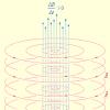
Cell tall-like receptor 9 ( toll-Like Receptor 9, TLR9) - one of the representatives of the "first line" of the body's immune response - specifically associates viral and bacterial DNA, forming characteristic M-shaped dimeric structures. The interaction with pathogenic DNA occurs due to the presence of a special component - cytosine-phosphate guanine (CPG) dinucleotide motif, which selectively communicates with the receptor in certain sites. The establishment of the crystal structure of the receptor-motive complex helped better understand the features of the work of this component of congenital immunity.
A detailed study of the features of the binding of the CPG-motive with the receptor showed that the immunostimulating activity of the dinucleotide depends on several important factors.
First And the most obvious of these is the number of non-generated cytosine-guanin sequences in the "hostile" DNA. The more CPG motifs are present in the nucleotide chain of bacteria, the greater the number of receptors will bind it.
Second A feature that managed to identify is a certain "pattern" of the motive expressed by the formula RR CG.Yy (Where FROM - cytosin, G. - Guanine, and R. and Y. - any purine and pyrimidine bases, respectively). It is noteworthy that inversion C and G leads to the formation of only the inactive monomer complex TLR9-CPG, while the formation of active implies dimer M-shaped Structure with recipe ratio: Ligand is 2: 2.
Third The factor is the processing of the receptor, which is necessary for the formation of a stoichiometric dimer. If the processing was not either he passed incorrectly, almost only monomeric forms were formed. The unbound Dimer TLR9 is the so-called z-loop consisting of leucine-rich sections ( lEUCINE-RICH REPEAT, LRR) (Fig. 1).
The CPG-Motive Binding Mechanism with the Recipe Site Authors of the Research Ophissed " molecular glue" The single-stranded DNA fragment is wrapped around the receptor, starting with the N-end of the protein molecule and covering several LRR sites. It is one nucleotide chain that can tightly facilitate the necessary sections of TLR9: when trying to use in experiments, the dual-stranded DNA receptor affinity decreased dramatically.
The CPG-motive itself, according to the above template, presented in the form of a hexamera, formed a complex system of hydrogen bonds with two dozen amino acids and additionally coordinated van der Wales interactions with a dozen residues. It is important that two DNA molecules have been subjected to a similar "attack from the flanks", since the associated receptor complex exists in the form of gomodimer (Fig. 2). Despite the abundance of amino acids connecting the CPG-motive, the mutation of some of them even separately can seriously reduce the "sticking" dinucleotide to the receptor.
What can be useful for the deep detection of the features of TLR9? Of course, the creation of targeted drugs for stimulation or, on the contrary, inhibiting the activity of these receptors. Violations in the work of the immune system (both in the direction of activation and suppression) underlie the set of infectious and autoimmune diseases. Knowledge of the structures and mechanisms of work of participants in congenital immunity will undoubtedly allow them to regulate and return the "knocked" parameters to normal.
Send your good work in the knowledge base is simple. Use the form below
Students, graduate students, young scientists who use the knowledge base in their studies and work will be very grateful to you.
Posted by http: // www. Allbest. RU /
Introduction
Tall-like receptors (TLR) are the main components of a congenital immunity system, which mediate the specific recognition of the evolutionary conservative molecular structures of pathogens (Pamp - Pathogen Associated Molecular Patterns). Tall-like receptors are presented on cells of different types - from epithelial to immunocompetent. As is known, when binding TLR with its own ligands, a number of adapter proteins and kinases are activated, which are involved in the induction of key pro-inflammatory factors. The result of such an induction is the development of both a congenital immune response as a result of increased expression of a number of anti-apoptotic proteins, pro-inflammatory cytokines, antibacterial proteins, and an acquired immune response through maturation of dendritic cells, antigen presentation, etc.
Due to its ability to strengthen the specific and nonspecific immune responses of the body, the agonists of tall-like receptors have found applications not only in the treatment of infectious diseases, but also as adjuvants in chemotherapy of various malignant neoplasms. However, it is currently described by fundamentally different TLR effects on the tumor. On the one hand, it was shown that TLR (and their ligands) can act as tumor growth suppressors, on the other hand, TLRs can stimulate tumor progression and affect the stability of tumor and chemotherapy. The presented review summarizes the data on the effect of TLR and their agonists on tumor growth, as well as the main mechanisms underlying such differences.
List of TLR contractions - Tall-like receptors; LPS - Lipopolysaccharide; NF-KB is a nuclear transcription factor KB; PRR - pattern recognizing receptors; Pamp - pathogen-associated molecular patterns; DAMP - molecular patterns associated with damage; IRF - interferonregulating factor, DCRNA - one-time dual-stranded ribonucleic acid; TNF-B - tumor necrosis factor B; IL - interleukin; IFN - Interferon; NK cells - natural killers; MirNNA - small interfering RNA; TGF - transforming growth factor.
1. History of opening
receiver immune antitumor pathogen
In 1985, in the study of various mutations at the flush-drosophila, the famous German biologist Christian Nyuslein-Folhard discovered larvae mutants with a underdeveloped ventral part of the body. Her immediate replica was "Das War Ja Toll!" ("This is a class!"). The epithet tall (cool) was later given to the corresponding gene as its name.
In 1996, it turned out that this gene is responsible not only for the dorgentral polarization in embryonic development, but also for the stability of drosophila to fungal infection. This opening of the French scientist Jules Hoffman was awarded the Nobel Prize of 2011. In 1997, Ruslan Medzhitov and Charles Jenauiz of the Yale University found a tall-like homologous gene in mammals (now he is called TLR4). It turned out that TLR4 causes activation of the Kappa-B NF-KB nuclear factor in the same way as, and interleukin-1. Finally, in 1998 it turned out that the ligand for the receptor is the component of the cell wall of gram-negative bacteria lipopolysaccharide.
2. TLR immune system
2.1 Structure TLR.
According to its structural organization, TLRs refer to the IL-1 receptor family (IL-1R). TLR is transmembrane proteins that are expressed on the cell surface and in subcellular compartments (such as endosome). The Localization of TLR is related to the type of ligand recognizable by him. Thus, TLR 1, 2, 4, 5, 6, binding structural bacterial components, are localized on the cell surface, while TLR 3, 7, 8, 9, recognizing predominantly virus-associated structures - nucleic acids (DCRNA, OCRNA, DNA) are in endosomes, where they interact with ligands after the deproteinization of virions.
The TLR structure is released by N-terminal leucinth (LRR) domain responsible for binding ligands, a transmembrane domain and a C-terminal intracellular signal domain (homologous IL-1R intracellular domain).
TLR is expressed in most types of human organism cells, including non-hymopoietic epithelial and endothelial cells. The amount of simultaneously expressed TLR and their combination are specific for each type of cells, and most of all TLR in cells of hematopoietic origin, such as macrophages, neutrophils, dendritic cells
At the moment, mammals identified 13 different TLRs, in humans - 10 and 12 in mice. TLR from the 1st to the 9th conservative in humans and mouse. However, there are differences. The gene encoding TLR10 is found only in humans, and TLR11 - both species, but functional only in mice.
The main feature of TLR, which distinguishes them from receptors of the acquired immunity (TI B-cell receptors), consists in their ability to recognize non-unique epitopes, and evolutionary conservative pathogen-associated molecular structures (Pamp), widely represented in all classes of microorganisms and viruses, regardless of their pathogenicity. PAMP recognition specificity is well studied in most TLRs, today Ligands TLR 1-9 and 11 (Fig. 1) are known. The biological role and the specificity of the TLR10 (person), 12 and 13 (mouse) remain unknown.
The most famous microbial ligands TLR:
bacterial lipopeptides, lipotheaic acid and peptidoglycans; Lipoarabidomananic myobacteria; The component of the cell wall of wintering mushrooms, which bind to TLR2, forming the heterodimers with TLR1, TLR6 and CD14;
LPS gram-negative bacteria, Ligand TLR4;
bacteria flavored components - flagllin, activating TLR5; Profiline-like structures of the simplest, binding to TLR11;
DNA (non-generated CPG sequences), recognizable TLR9;
dCRNA - Ligand TLR3;
oCRNA - Ligands TLR7 and TLR8.
Recently it was shown that TLRs can be activated by many endogenous molecules - allarminamines (hyaluronic acid, heat shock proteins, etc.), which appear in the destruction of tissues. These heterogeneous in nature and structure of the connection (Pamp and Allarmines) recognizable by TLR are currently united in one family, called DAMP (Damage Associated Molecular Patterns)
2.2 TLR interaction with its own ligands
Now, from the description of the structure and functions of the TLR, we turn to events unfolding after their binding with our own ligands.
The binding of the ligand C TLR initiates the signal cascade, originating from cytoplasmic TLR TIR-domains. TIR-domain signal via MYD88 adapter molecules (MYELOID DIFFERENTIATION FACTOR 88), TIRAP (TIR domain-containing adapters), TiCam1 (TRIF), Ticam2 (TIR-Containing AdapteR Molecule) is transmitted to the corresponding kinases (TAK, IKK, TBK, MAPK, JNKS, P38, ERK, AKT, etc.), which differentially activate transcription factors (NF-KB, AP-1 and IRF), responsible for expression of various pro-inflammatory and antimicrobial factors. In this case, all TLRs, except for TLR3, transmit a signal to kinase using MYD88. TLR3 transmits a signal via Ticam1, A TLR4 and through MyD88, and through Ticam1.
The activation of one or another factor is determined by the TLR type, from which the signal is transmitted. So, almost all TLR (TLR2 and its coreceptors - TLR1 and TLR6, as well as TLR4-9, TLR11), binding to its own ligands, are able to activate NF-KB - one of the main factors governing the expression of such pro-inflammatory cytokines as IL-1 , -6, -8, etc. To activate another family of pro-inflammatory transcription factors, the IRF leads the signal transmission through TLR3, 4, 7-9. The signals transmitted through TLR3 or TLR4 lead to IRF3 activation, which regulates the expression of IFN-B and is considered a critical component of antiviral immune reactions. The transmission of signals through TLR7-9 leads to the activation of IRF5 and IRF7 and expression of IFN-B, which also plays a vital role in antiviral protection. Alarm through TLR2 or TLR5 does not lead to activation of the factors of the IRF family.
Thus, the interaction of TLR of definitely type with its own ligand initiates the launch of the signal cascade, which leads to activation of expression of a specific combination of genes (cytokines, antimicrobial molecules, etc.). However, currently a lot of activation of TLR-dependent signaling pathways and in the development of subsequent effects remains incomprehensible. In the affordable scientific literature there are no data that characterize full transcriptomic and proteoma changes that occur in response to the activation of certain TLRs.
3. TLR functions
TLRs on the body running in the body are related to the PRR family, which mediate the specific recognition of the evolutionary conservative structures of pathogens (Pamp - Pathogen Associated Molecular Patterns). Binding to RAMR, TLR activates the system of congenital immunity and largely determine the development of adaptive immunity. The most conservative role of TLR is the activation of antimicrobial immunity in the skin, mucous membranes of the respiratory, gastrointestinal and urogenital tract.
TLR recognize microbial molecules, which leads to the development of inflammatory reactions caused by the activation of the NF-KB factor, which regulates the expression of pro-inflammatory cytokines (TNF-B, IL-1, IL-6, etc.) and Chemokins (MCP-1, MCP-3 , GMCSF, etc.).
TLR is involved in transcriptional and post-translational regulation (proteolytic splitting and secretion) of antimicrobial factors such as defense (b and c), phospholipase A2, lysozyme, etc. TLR enhance the absorption of microorganisms by phagocytes and optimize their inactivation, adjusting the emission of peroxidant radicals and nitrogen oxide.
It is known that TLRs on the surface of endothelial cells indirectly ensure the migration of leukocytes into the focus of inflammation, stimulating the expression of the leukocyte adhesion molecules - e-selectin and ICAM-1.
TLR stimulation directly leads to an increase in interferon products (IFN) -B / in both stromal and hematopoietic cells, which is important to protect the organism from viral and some bacterial infections. Moreover, it has recently been established that TLR, activating a series of molecules (FADD, Caspanis 8, proteincinase R (PKR)) or stimulating the expression of IFN-b / c, can induce the development of apoptosis - an important mechanism that protects cells from pathogenic microorganisms.
It is shown that TLR play a central role in regulating an adaptive immune response. Thus, TLR-dependent activation of professional antigen-representing dendritic cells is a determining point in several fundamental for the development of adaptive immunity of processes: activation of mature T cells; processing and presentation of microbial antigens; increasing the expression of knitimulatory molecules (CD80, CD86) required to activate naive CD4 + -TT cells; suppressing regulatory T cells by product IL-6. It is also known that tlper-dependent activation is important for proliferation and ripening in cells during infection.
Thus, the TLR is performed in the body an important role that consists in the development of inflammatory reactions (activation of congenital immunity) in response to entering the body of various pathogens (simplest, fungi, bacteria, viruses). Moreover, according to modern ideas, pathogen recognition by TLR is a key point in the formation of the second protection line - adaptive immunity. It is also shown that TLR takes part in the normal functioning of the intestine, they are involved in the development of autoimmune diseases (systemic lupus), arthritis, atherosclerosis, etc. Recently, data has been obtained that show that TLRs are able to activate antitumor immunity or, on the contrary, stimulate tumor Progression.
3.1 Tumor Activity TLR
Many TLR agonists are currently undergoing clinical trials as antitumor agents. So, natural (OCRNA) and synthetic (imiquine) agonists TLR7 and 8 showed high activity against chronic lymphocytic leukemia and skin tumors. Ligand TLR9 - CPG is capable of suppressing the growth of lymph, brain tumors, kidneys, leather. And the ligand TLR3 - POLY (IC) has a pro -appopototic effect not only in relation to tumor cells, but also of environment cells (for example, endothelium).
It is shown that the TLR4 agonists - LPS gram-negative bacteria and OK-432 (the preparation from streptococci Group a), have high antitumor activity with intratumolar administration. However, with systemic administration, both drugs (LPS and OK432) did not have the ability to block tumor growth. Currently, the OK-432 drug passes the second stage of clinical trials, as a means against colorectal tumors and lung cancer. It is also shown that OM-174, the TLR2 / 4 chemical agonist is capable of suppressing the progression of melanoma and increase the survival of experimental animals when administered with cyclophosphamide. In these experiments, the TLR2 / 4 agonists induce the TNF-B secretion and expression of inducible NO synthase. As it is known, no is able to induce apoptosis in tumor cells resistant to chemotherapy, and thereby increase the life expectancy of mice. Another known antitumor drug microbial origin, activating TLR-dependent response (TLR2, 4, 9), - BCG. This drug has been relatively successfully used in the treatment of urinary tumor therapy.
In general, it should be noted that at present various TLR agonists undergo clinical trials as a means against tumors of various origin.
One of the main mechanisms of antitumor activity TLR consists in their ability to stimulate the development of a tumor-specific immune response. So, activation TLR:
1) stimulates (directly or indirectly) migration into a nk cell tumor, cytotoxic T cells and T-helpers of the i-th type, which cause lysis of tumor cells using various effector mechanisms (secretion of perforins, grazes, IFN-g, etc. );
2) leads to the secretion of IFN I type (IFN-B, B). Another probable TLR antitumor activity mechanism is the possibility of TLR-dependent transition of the tumor-fluent type of macrophages (M2) into the tumor-absorbing type M1. Macrophages of the M2 type are characterized by the expression of cytokines such as TGF-B and IL-10, the components necessary for repairing and remodeling tissues. TGF-B stimulates the proliferation of tumor cells, IL-10 sends the development of an immune response toward TH2, thereby blocking the development of cellular antitumor immunity. Macrophages type M1, on the contrary, express IL-1, -6, -12, TNF-B, IFN-G and stimulate the development of antitumor cellular (TH1) immune response.
3.2 Tumor Femulating activity TLR
As is known, chronic infections and inflammation are essential factors that stimulate the development of malignant neoplasms. In particular, the stomach cancer may be associated with chronic inflammation caused by such a pathogen as Helicobacter Pylori, and the chronic inflammation of the digestive tract is often associated with the development of colon cancer. Moreover, it is shown that the use of non-steroidal anti-inflammatory drugs can reduce the risk of developing certain types of malignant neoplasms.
TLR serve as a key link of the system of congenital immunity and animals, they participate in the development of inflammatory reactions when cells with different pathogens. Currently, the role of the TLR in the development and progression of tumors of various origin is actively being studied. TLR can be involved in the development and stimulation of tumor formation by several mechanisms.
One of the most important factors caused by the relationship of chronic inflammation and tumor formation is NF-KB. This factor is constitutively activated by more than 90% of human tumors, including acute and chronic myeloid leukemia, prostate cancer, multiple ate, malignant hepatom (liver cancer), etc.
In this regard, agents capable of activating NF-KB can directly participate in the development and progression of the tumor. As is known, the interaction of pathogens with TLR on the cell surface leads to the activation of NF-KB and the expression of NF-KB-dependent genes, which necessitates the participation of TLR in the stimulation of carcinogenesis. Activation of NF-KB leads to an increase in the production of cytokines IL-1, IL-2, IL-6, IL-10, TNF-B; migration of cells of the immune system to the place of inflammation as a result of improving the products of chemokines; "Maintaining" chronic inflammation; Increased products of anti-apoptotic factors, etc. These properties can ensure the survival and progression of the tumor due to the suppression of apoptosis and cytotoxicity, as well as induction of angiogenesis.
Currently, it is known that the TLR level is raised in the cells of various tumors, and in mice with knocuite TLR genes reduced the frequency of the formation of tumors inducing. Moreover, an increase in the expression of TLR on the surface of the prostate tumor cells or head tumors and neck can stimulate their proliferation.
Huang et al. It has shown that Listeria Monocytogenes has a direct tumor-fluent action associated with its ability to activate TLR2-dependent signaling pathways in ovarian cancer cells. Moreover, TLR2-dependent activation of NF-KB, caused by L. monocytogenes, led to an increase in the stability of tumor cells to the action of chemotherapeutic drugs.
The relationship of TLR2 with tumor progress is confirmed in another independent study in which Karin et al. Proved the key role of this receptor in metastasis the lung cancer. It turned out that in mice with a knockout of the TLR2 gene, metastasis and the progression of tumors occurs significantly slower than that of wild type mice. The key role in the progression of lung cancer was played by myeloid cells expressing TNF-B in response to their versican stimulation (proteoglycan of extracellular matrix, the Ligand TLR2, the level of which is raised in tumor cells of many types). Our studies also studied the role of TLR2 in tumor progression. In particular, it turned out that mycoplasma infection (MyCoplasma ARGININI) or the addition of structural components (Lamb) of this pathogen to cells expressing TLR2 leads to the suppression of apoptosis in them, as well as to strengthen the tumor growth in in vivo. Thus, it is shown that the TLR can have a mediated tumor distribution effect through the cells of myeloid series.
Similar data were obtained for another representative of the TLR4 family. Systemic (intravenous) administration of the ligand of this receptor - LPS stimulated the migration of tumor cells (breast adenocarcinoma) and increased their invasiveness, and also stimulated angiogenesis in tumors. Similar results were obtained on another model - the intestinal adenocarcinoma: LPS increased the survival rate of the tumor cells, stimulated their proliferation, and in the intraperitoneal administration, increased metastasis. Moreover, Huang et al. It was shown that tumor cells expressing TLR4 cause a significantly more aggressive course of the disease (reducing the life of animals) compared to mice of the isogenic line, in which TLR4 is inactivated by a specific mrynk. The data obtained allowed us to assume that endogenous ligands (heat shock proteins; in-defense; endogenous LPS, thrown from the intestine, can be influenced by the progression of TLR4-positive tumors; which is partly reminding the situation with the TLR2 tumoring effect and its ligand of endogenous origin - versicane.
However, the data illustrating the TLR tumor power supply is obtained not only for TLR2 and 4. It is known that the increased expression of TLR5 and TLR9 on the cells of the cervical epithelium can be associated with the progression of the cervical cancer. The high level of expression TLR9 is detected in clinical samples of lung cancer and in tumor cell lines. In these cells, the stimulation TLR9 with specific agonists led to an increase in production of tumor-associated cytokines. The level of TLR9 is also elevated on the surface of the human prostate gland tumor cell. The treatment of such CPG-oligodeoxinucleotide cells (ODN-CPG) or bacterial DNAs serving ligands for TLR9 has contributed to an increase in the invasion of tumor cells. An increase in the invasion of tumor cells as a result of activation of TLR9 can be considered as a new mechanism by which chronic infections can stimulate the growth of prostate tumor cells.
However, the ability to stimulate carcinogenesis through interaction with TLR has not only different infectious agents and their structural components. As you know, the ligands for TLR also will also serve DAMP - nuclear and cytoplasmic cells of cells undergoing necrosis. Released from damaged DAMP cells can be recognized by various TLRs on the surface of immune cells, and the subsequent activation of Tlred-dependent signals can lead to the suppression of an antitumor immune response and, as a result, to stimulate the progression of the tumor.
To such molecules with a potential tumor distributor include: heat shock proteins (HSP60, 70), APP and urinary acid, CA2 +-modulating proteins (S100), HMGB1 protein and nucleic acids, of which the DNA-binding protein of HMGB1 is most well studied . Elevated as a result of damage to the cells of the HMGB1 protein activates the immune system through the interaction with TLR. The cultures of cells showed that the HMGB1 protein stimulates the growth of melanoma cells, breast cancer, colon, pancreas and prostate gland. HMGB1 is able to activate TLR2 and TLR4 on the tumor cells and cells of the immune system and, as a result, induce tumor progression and metastasis.
It is shown that the melanoma cells have increased expression of such DAMP, as the S100 family proteins that can stimulate the growth and melanoma cells themselves, and peripheral blood lymphocytes, acting as an autocreen tumor growth factor. S100A4 protein serving ligand for TLR stimulates metastasis of breast cancer cells, and its increased expression is an indicator of a bad forecast. Despite the relationship of S100A4 with metastasis, this protein can be expressed by macrophages, lymphocytes and fibroblasts. Recent studies have shown that S100A8 and S100A9 proteins produced by the primary tumor are capable of activating whey amyloid A (SAA) 3 in pulmonary tissues and thereby create conditions for the formation of a metastatic niche. SAA3 serves as a ligand for TLR4 on endothelial cells of light and macrophages. The activation of TLR4 facilitates the migration of tumor cells from the primary focus to the tissue of the lungs by forming a microenvironment that contributes to the growth of the tumor. Thus, the suppression of the S100-TLR4 signaling path can effectively counteract the formation of metastases in light.
Summing up the effects described, it can be concluded that the TLR ability, on the one hand, directly or indirectly participate in tumor progression, and on the other, to increase the stability of tumor cells to pro-apoptotic effects.
The data presented shows that TLR tumoriformulating effects and their ligands have a complex mechanism that needs to be studied in more detail. However, despite the complexity of this issue, several key moments can be distinguished that determine the TLR tumor fimulting effect:
1) The interaction of TLR with its own ligands induces the activation of the transcriptional factor NF-KB and, as a result, increasing the products of various pro-inflammatory cytokines (IL-6, MCP-1, MIF, GROD, etc.), as well as a number of anti-apoptotic proteins, thereby contributing direct or indirect tumor distribution action;
2) TLR-dependent activation of myeloid cells and their predecessors, apparently, is a determining factor in the formation of metastases. In a series of independent work, it is shown that myeloid cells migrating from bone marrow (in response to endogenous stimulation) in tissue play a key role in the formation of metastatic niche. Since it is known that endogenous (versican, fibronectin, etc.) and exogenous (microbial origin) of the TLR ligands are capable of, on the one hand, stimulate myeloid cells and their predecessors, and on the other - to increase the metastatic potential of the tumor, then it is possible to assume the existence with a high probability the relationship between TLR-dependent activation of myeloid cells and their subsequent participation in metastasis;
3) Activation of TLR can stimulate angiogenesis through antigenic factors such as IL-8, the growth factor of vascular endothelium (VEGF) and matrix metalloproteinases (MMP), as well as enhance the adhesive and invasive properties of tumor cells, along with an increase in vessel permeability.
Posted on AllBest.ru.
...Similar documents
Opening of communication between the immune and nervous systems of the body. Glutamate receptors in the nervous system and their purpose. Molecular reactions of the activated neuron. Causes and consequences of NMDA receptor neurotoxicity. Signing alive neurons.
abstract, added 05/26/2010
Cytokines and their cell receptors. Phagocytosis as an important component of antimicrobial protection. The choice of effector mechanisms of cellular immunity. Network interactions of cytokines. Reactions aimed at eliminating viruses of organism cells infected with viruses.
abstract, added 09/28/2009
Characteristic of dopamine, its properties and functions in the human body; Ensuring cognitive activities. Classification of dopamine receptors: species, localization. Description of the mechanisms of action and side effects of various dopaminergic agents.
presentation, added 06/15/2015
The role of obese cells in the regulation of the organism homeostasis. Localization of fat cells, their mediators. Secretion of mediators and their functions. The main types of fat cells. Receptors and ligands, effects of mediators. Participation of fat cells in pathological processes.
presentation, added 01/16/2014
Morphological manifestations of the development of inflammatory response of the body on tuberculosis infection. The study of enzymatic reactions, on the activity of which depends on the functional state of all organs and the body as a whole. The role of connective tissue cells.
abstract, added 15.09.2010
Jenner as the founder of immunite doctrine. Nonspecific cellular and humoral protective mechanisms. Specific immune systems. Immunity organs: Milk iron (thymus), bone marrow, lymph nodes, lymphoid fabric of the spleen.
abstract, added 04.02.2010
Substances that can cause allergies. Immune body reactions. The formation of antigenspecific clones. Slow-type hypersensitivity reactions. Stage of pathophysiological changes. The main methods of treating allergic diseases.
abstract, added 07.10.2013
Evaluation of the effectiveness and safety of treatment arterial hypertension In patients APE inhibitors, receptor blockers to angiotensin, diuretics. Familiarization with the results of therapy with leaser, losartan, verapamil, betaxolol, hypothiazide.
abstract, added 24.07.2014
Determination of the concept of "endometriosis". Participation in the mechanism of the disease of cell enzymes, receptors to hormones, as well as gene mutations. Etiology, pathogenesis, classification and clinical picture of genital endometriosis. Diagnosis and treatment of the disease.
presentation, added 09/23/2014
Bioelectric phenomena in nerve cells. Characteristics of receptors, their types and specificity, the concepts of "neuromediator", "messenger", structure and mechanism of their action. The effect of pharmacological agents in the treatment of diseases of the central nervous system.